

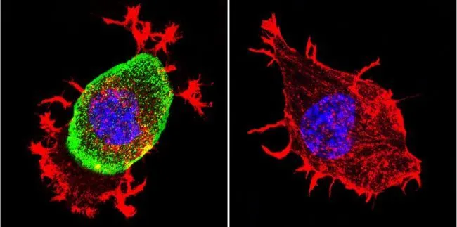
ICC/IF analysis of Neuro-2a Cells using GTX22803 AChE antibody [HR2]. Cells were probed without (right) or with(left) an antibody. Green : Primary antibody Blue : Nuclei Red : Actin Fixation : formaldehyde Dilution : 1:200 overnight at 4oC
![IHC-P analysis of human rectum tissue using GTX22803 AChE antibody [HR2]. Left : Primary antibody Right : Negative control without primary antibody Antigen retrieval : heat induced antigen retrieval was performed using 10mM sodium citrate (pH6.0) buffer, microwaved for 8-15 minutes Dilution : 1:20](https://www.genetex.com/upload/website/prouct_img/normal/GTX22803/GTX22803_1132_IHC-P_w_23060620_807.webp "IHC-P analysis of human rectum tissue using GTX22803 AChE antibody [HR2]. Left : Primary antibody Right : Negative control without primary antibody Antigen retrieval : heat induced antigen retrieval was performed using 10mM sodium citrate (pH6.0) buffer, microwaved for 8-15 minutes Dilution : 1:20")
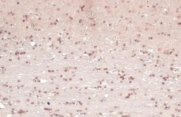
![IHC-P analysis of human Brain tissue using GTX22803 AChE antibody [HR2]. Left : Primary antibody Right : Negative control without primary antibody Antigen retrieval : heat induced antigen retrieval was performed using 10mM sodium citrate (pH6.0) buffer, microwaved for 8-15 minutes Dilution : 1:200](https://www.genetex.com/upload/website/prouct_img/normal/GTX22803/GTX22803_1130_IHC-P_w_23060620_277.webp "IHC-P analysis of human Brain tissue using GTX22803 AChE antibody [HR2]. Left : Primary antibody Right : Negative control without primary antibody Antigen retrieval : heat induced antigen retrieval was performed using 10mM sodium citrate (pH6.0) buffer, microwaved for 8-15 minutes Dilution : 1:200")
![ICC/IF analysis of HeLa cells using GTX22803 AChE antibody [HR2]. Cells were probed without (right) or with(left) an antibody. Green : Primary antibody Blue : Nuclei Red : Actin Fixation : formaldehyde Dilution : 1:200 overnight at 4oC](https://www.genetex.com/upload/website/prouct_img/normal/GTX22803/GTX22803_475_ICC-IF_w_23060620_926.webp "ICC/IF analysis of HeLa cells using GTX22803 AChE antibody [HR2]. Cells were probed without (right) or with(left) an antibody. Green : Primary antibody Blue : Nuclei Red : Actin Fixation : formaldehyde Dilution : 1:200 overnight at 4oC")
![ICC/IF analysis of U251 cells using GTX22803 AChE antibody [HR2]. Cells were probed without (right) or with(left) an antibody. Green : Primary antibody Blue : Nuclei Red : Actin Fixation : formaldehyde Dilution : 1:200 overnight at 4oC](https://www.genetex.com/upload/website/prouct_img/normal/GTX22803/GTX22803_477_ICC-IF_w_23060620_199.webp "ICC/IF analysis of U251 cells using GTX22803 AChE antibody [HR2]. Cells were probed without (right) or with(left) an antibody. Green : Primary antibody Blue : Nuclei Red : Actin Fixation : formaldehyde Dilution : 1:200 overnight at 4oC")
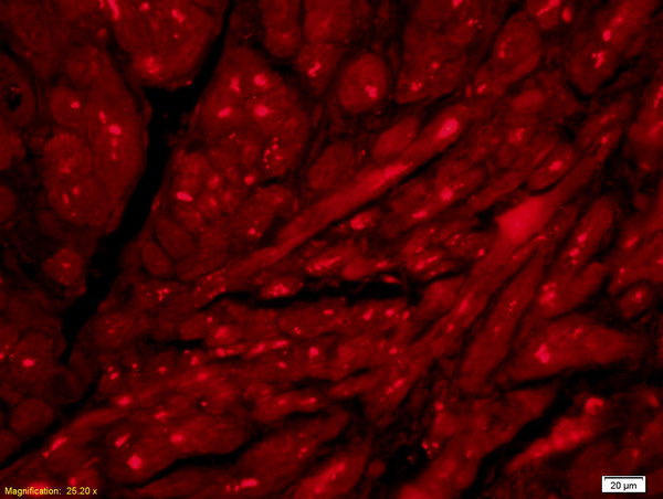
![IHC-P analysis of human cerebellum tissue using GTX22803 AChE antibody [HR2]. Left : Primary antibody Right : Negative control without primary antibody Antigen retrieval : heat induced antigen retrieval was performed using 10mM sodium citrate (pH6.0) buffer, microwaved for 8-15 minutes Dilution : 1:50](https://www.genetex.com/upload/website/prouct_img/normal/GTX22803/GTX22803_1131_IHC-P_w_23060620_833.webp "IHC-P analysis of human cerebellum tissue using GTX22803 AChE antibody [HR2]. Left : Primary antibody Right : Negative control without primary antibody Antigen retrieval : heat induced antigen retrieval was performed using 10mM sodium citrate (pH6.0) buffer, microwaved for 8-15 minutes Dilution : 1:50")
ICC/IF analysis of Neuro-2a Cells using GTX22803 AChE antibody [HR2]. Cells were probed without (right) or with(left) an antibody. Green : Primary antibody Blue : Nuclei Red : Actin Fixation : formaldehyde Dilution : 1:200 overnight at 4oC
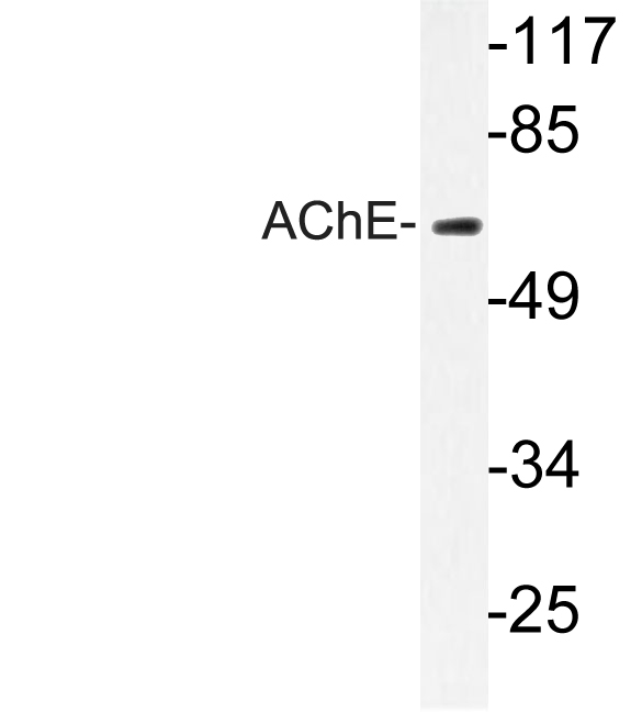
AChE antibody [HR2]
GTX22803
ApplicationsImmunoFluorescence, ImmunoPrecipitation, Western Blot, ELISA, ImmunoCytoChemistry, ImmunoHistoChemistry, ImmunoHistoChemistry Frozen, ImmunoHistoChemistry Paraffin
Product group Antibodies
ReactivityBovine, Feline, Guinea Pig, Human, Mouse, Rabbit
TargetACHE
Overview
- SupplierGeneTex
- Product NameAChE antibody [HR2]
- Delivery Days Customer9
- Application Supplier NoteICC/IF: 1:100-1:1000. IHC-Fr: 1:50. *Optimal dilutions/concentrations should be determined by the researcher.Not tested in other applications.
- ApplicationsImmunoFluorescence, ImmunoPrecipitation, Western Blot, ELISA, ImmunoCytoChemistry, ImmunoHistoChemistry, ImmunoHistoChemistry Frozen, ImmunoHistoChemistry Paraffin
- CertificationResearch Use Only
- ClonalityMonoclonal
- Clone IDHR2
- Concentration1 mg/ml
- ConjugateUnconjugated
- Gene ID43
- Target nameACHE
- Target descriptionacetylcholinesterase (Yt blood group)
- Target synonymsACEE, ARACHE, N-ACHE, YT, acetylcholinesterase, Yt blood group, acetylcholinesterase (Cartwright blood group), apoptosis-related acetylcholinesterase
- HostMouse
- IsotypeIgG2b
- Protein IDP22303
- Protein NameAcetylcholinesterase
- Scientific DescriptionAcetylcholinesterase hydrolyzes the neurotransmitter, acetylcholine at neuromuscular junctions and brain cholinergic synapses, and thus terminates signal transmission. It is also found on the red blood cell membranes, where it constitutes the Yt blood group antigen. Acetylcholinesterase exists in multiple molecular forms which possess similar catalytic properties, but differ in their oligomeric assembly and mode of cell attachment to the cell surface. It is encoded by the single ACHE gene, and the structural diversity in the gene products arises from alternative mRNA splicing, and post-translational associations of catalytic and structural subunits. The major form of acetylcholinesterase found in brain, muscle and other tissues is the hydrophilic species, which forms disulfide-linked oligomers with collagenous, or lipid-containing structural subunits. The other, alternatively spliced form, expressed primarily in the erythroid tissues, differs at the C-terminal end, and contains a cleavable hydrophobic peptide with a GPI-anchor site. It associates with the membranes through the phosphoinositide (PI) moieties added post-translationally. [provided by RefSeq, Jul 2008]
- ReactivityBovine, Feline, Guinea Pig, Human, Mouse, Rabbit
- Storage Instruction-20°C or -80°C,2°C to 8°C
- UNSPSC12352203
References
- Jean L, Brimijoin S, Vaux DJ. In vivo localization of human acetylcholinesterase-derived species in a β-sheet conformation at the core of senile plaques in Alzheimer's disease. J Biol Chem. 2019,294(16):6253-6272. doi: 10.1074/jbc.RA118.006230Read this paper
- Dafferner AJ, Schopfer LM, Xiao G, et al. Immunopurification of Acetylcholinesterase from Red Blood Cells for Detection of Nerve Agent Exposure. Chem Res Toxicol. 2017,30(10):1897-1910. doi: 10.1021/acs.chemrestox.7b00209Read this paper
Datasheet
Related products
Product group Antibodies

Anti-acetylcholinesterase [1G]AB04029-1.1
ApplicationsELISA
ReactivityHuman
TargetACHE
- SizePrice
Product group Antibodies
Anti-ACHE Antibody144-02806
ApplicationsWestern Blot, ImmunoHistoChemistry
ReactivityHuman, Mouse, Rat
TargetACHE
- SizePrice
Product group Antibodies
References
AChE antibodyGTX101648
ApplicationsImmunoFluorescence, Western Blot, ImmunoCytoChemistry, ImmunoHistoChemistry, ImmunoHistoChemistry Frozen, ImmunoHistoChemistry Paraffin
ReactivityHuman, Mouse, Rat
TargetACHE
- SizePrice
![AChE antibody [HL1102] detects AChE protein at dendrite by immunofluorescent analysis. Sample: DIV9 rat E18 primary cortical neuron and glia cells were fixed in 4% paraformaldehyde at RT for 15 min. Green: AChE stained by AChE antibody [HL1102] (GTX636298) diluted at 1:250. Red: Tau, an axon marker, stained by Tau antibody [GT287] (GTX634809) diluted at 1:500. Blue: Fluoroshield with DAPI (GTX30920).](https://www.genetex.com/upload/website/prouct_img/normal/GTX636298/GTX636298_44410_20221209_ICC_IF_R_22122018_829.webp)
Product group Antibodies
AChE antibody [HL1102]GTX636298
ApplicationsImmunoFluorescence, Western Blot, ImmunoCytoChemistry, ImmunoHistoChemistry, ImmunoHistoChemistry Paraffin
ReactivityFeline, Human, Mouse, Rat
TargetACHE
- SizePrice
Product group Antibodies
References
ACHE Polyclonal AntibodyBS-2511R
ApplicationsImmunoFluorescence, Western Blot, ELISA, ImmunoCytoChemistry, ImmunoHistoChemistry, ImmunoHistoChemistry Frozen, ImmunoHistoChemistry Paraffin
ReactivityBovine, Canine, Equine, Human, Mouse, Rat
TargetACHE
- SizePrice
Product group Antibodies
ACHE AntibodyCSB-PA010202
ApplicationsWestern Blot, ELISA, ImmunoHistoChemistry
ReactivityHuman, Mouse, Rat
TargetACHE
- SizePrice
Product group Antibodies
AChE antibody, C-termGTX89580
ApplicationsFlow Cytometry, ImmunoFluorescence, Western Blot, ImmunoCytoChemistry
ReactivityHuman, Rat
TargetACHE
- SizePrice
Product group Antibodies
Anti-AChE AntibodyA96247
ApplicationsWestern Blot, ELISA, ImmunoHistoChemistry
ReactivityHuman, Mouse, Rat
- SizePrice