


Cross-reactivity assessment of ACP5 Mouse Recombinant Antibody AE00100 (1ug/ml) on CDI’s Protein Array containing more than 19,000 full-length human proteins.

 and intact heavy and light chains at 50kDa and 25kDa resp. (R).")
Cross-reactivity assessment of ACP5 Mouse Recombinant Antibody AE00100 (1ug/ml) on CDI’s Protein Array containing more than 19,000 full-length human proteins.
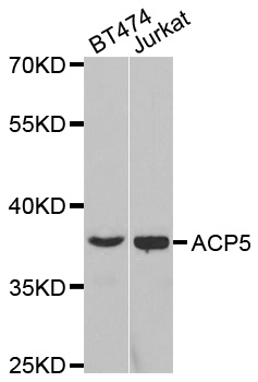
ACP5 Recombinant Antibody AE00100
AE00100
TargetACP5
Product group Antibodies
Overview
- SupplierAeonian Biotech
- Product NameACP5 Recombinant Antibody AE00100
- Delivery Days Customer9
- Applications SupplierIHC, PA
- CertificationResearch Use Only
- ClonalityRecombinant
- Clone IDrACP5/1070
- ConjugateUnconjugated
- Gene ID54
- Target nameACP5
- Target descriptionacid phosphatase 5, tartrate resistant
- Target synonymsHPAP, TRACP5a, TRACP5b, TRAP, TRAcP, TrATPase, tartrate-resistant acid phosphatase type 5, human purple acid phosphatase, tartrate-resistant acid ATPase, tartrate-resistant acid phosphatase 5a, tartrate-resistant acid phosphatase 5b
- HostMouse
- IsotypeIgG1 kappa
- Protein IDP13686
- Protein NameTartrate-resistant acid phosphatase type 5
- Scientific DescriptionACP5 Recombinant Antibody AE00100
- Shelf life instructionIntegrity warranted for 24 months after purchase when handled and stored according to instructions, see below.
- Reactivity SupplierHuman
- Storage Instruction2-8°C
- UNSPSC12352203
Datasheet
MSDS
Related products
Product group Antibodies
ApplicationsImmunoPrecipitation, Western Blot, ImmunoCytoChemistry, ImmunoHistoChemistry
ReactivityMouse, Rat
TargetACP5
- SizePrice
Product group Antibodies
Anti-ACP5 Antibody, Rabbit Polyclonal100741-T32
ApplicationsWestern Blot
ReactivityHuman
- SizePrice
Product group Antibodies
Anti-ACP5 Antibody144-02528
ApplicationsImmunoFluorescence, Western Blot
ReactivityHuman, Mouse
TargetACP5
- SizePrice
Product group Antibodies
Anti-ACP5 AntibodyA35524
ApplicationsImmunoFluorescence, Western Blot, ImmunoHistoChemistry
ReactivityHuman, Mouse, Rat
- SizePrice
Product group Antibodies
TRAP antibody [ZM174]GTX01842
ApplicationsImmunoHistoChemistry, ImmunoHistoChemistry Paraffin
ReactivityHuman
TargetACP5
- SizePrice
Product group Antibodies
ACP5 AntibodyCSB-PA001178GA01HU
ApplicationsELISA, ImmunoHistoChemistry
ReactivityHuman, Mouse, Rat
TargetACP5
- SizePrice
![IHC-P analysis of human spleen tissue using GTX35145 TRAP antibody [ACP5/1070].](https://www.genetex.com/upload/website/prouct_img/normal/GTX35145/GTX35145_20200115_IHC-P_1173_w_23060801_480.webp)
Product group Antibodies
TRAP antibody [ACP5/1070]GTX35145
ApplicationsImmunoHistoChemistry, ImmunoHistoChemistry Paraffin
ReactivityHuman, Mouse, Rat
TargetACP5
- SizePrice
![IHC-P analysis of human spleen tissue section using GTX02582 TRAP antibody [ACP5/2336R].](https://www.genetex.com/upload/website/prouct_img/normal/GTX02582/GTX02582_20210319_IHC-P_w_23053122_289.webp)
Product group Antibodies
TRAP antibody [ACP5/2336R]GTX02582
ApplicationsImmunoHistoChemistry, ImmunoHistoChemistry Paraffin
ReactivityHuman, Mouse, Rat
TargetACP5
- SizePrice