

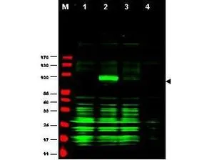
Western blot using Affinity Purified anti-Ajuba antibody shows detection of Ajuba-RFP fusion protein in cell lysates (arrow-head). Lanes correspond to 1) vector only trans-fection, 2) human Ajuba-RFP, 3) mouse Ajuba-RFP, and 4) mock transfection. Approximately 50 μg of each lysate was loaded per lane for SDS-PAGE followed by transfer onto nitrocellulose and reaction with a 1:1,700 dilution of anti-Ajuba antibody.
. Lanes correspond to 1) HeLa nuclear extract, and 2) HeLa, 3) A431, 4) Jurkat and 5) 293 whole cell lysates. Immunoprecipitation of Ajuba followed by western blotting may result in cleaner background staining. Approximately 5 μg of each preparation was run on a SDS-PAGE and transferred onto nitrocellulose followed by reaction with a 1:500 dilution of anti-Ajuba antibody. Detection occurred using a 1:5,000 dilution of HRP-labeled Donkey anti-Rabbit IgG for 1 hour at room temperature. A chemiluminescence system was used for signal detection (Roche) using a 60-sec exposure time.")

Western blot using Affinity Purified anti-Ajuba antibody shows detection of Ajuba-RFP fusion protein in cell lysates (arrow-head). Lanes correspond to 1) vector only trans-fection, 2) human Ajuba-RFP, 3) mouse Ajuba-RFP, and 4) mock transfection. Approximately 50 μg of each lysate was loaded per lane for SDS-PAGE followed by transfer onto nitrocellulose and reaction with a 1:1,700 dilution of anti-Ajuba antibody.
Ajuba antibody
GTX48743
ApplicationsImmunoPrecipitation, Western Blot, ELISA
Product group Antibodies
ReactivityHuman
TargetAJUBA
Overview
- SupplierGeneTex
- Product NameAjuba antibody
- Delivery Days Customer9
- Application Supplier NoteWB: 1:500-1:2500. IP: 1:100. ELISA: 1:20000-1:80000. *Optimal dilutions/concentrations should be determined by the researcher.Not tested in other applications.
- ApplicationsImmunoPrecipitation, Western Blot, ELISA
- CertificationResearch Use Only
- ClonalityPolyclonal
- Concentration1.67 mg/ml
- ConjugateUnconjugated
- Gene ID84962
- Target nameAJUBA
- Target descriptionajuba LIM protein
- Target synonymsJUB, LIM domain-containing protein ajuba, jub, ajuba homolog
- HostRabbit
- IsotypeIgG
- Protein IDQ96IF1
- Protein NameLIM domain-containing protein ajuba
- Scientific DescriptionHuman Ajuba (also called JUB protein and ajuba homolog isoform 1) is a LIM domain protein suggested to bind and regulate the activity of Aurora A. Aurora A, which is involved in cell cycle regulation, is upregulated during mitosis, localizing to the centrosomes and microtubule regions proximal to the centrosomes.
- ReactivityHuman
- Storage Instruction-20°C or -80°C,2°C to 8°C
- UNSPSC41116161
Datasheet
Related products
Product group Antibodies

JUB / Ajuba Antibody (aa75-125)LS-C762387
ApplicationsImmunoPrecipitation, Western Blot
ReactivityHuman
TargetAJUBA
- SizePrice

Product group Antibodies
Anti-AJUBA AntibodyHPA006171
ApplicationsWestern Blot, ImmunoCytoChemistry, ImmunoHistoChemistry
ReactivityHuman
TargetAJUBA
- SizePrice
Product group Antibodies
AJUBA AntibodyCSB-PA836229NA01HU
ApplicationsELISA, ImmunoHistoChemistry
ReactivityHuman
TargetAJUBA
- SizePrice
Product group Antibodies
Ajuba antibodyGTX134670
ApplicationsImmunoFluorescence, Western Blot, ImmunoCytoChemistry
ReactivityHuman
TargetAJUBA
- SizePrice