

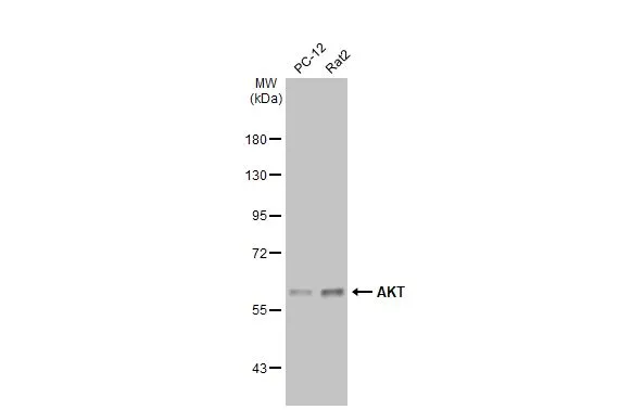
Various whole cell extracts (30 μg) were separated by 7.5% SDS-PAGE, and the membrane was blotted with AKT antibody [N3C2], Internal (GTX121937) diluted at 1:3000. The HRP-conjugated anti-rabbit IgG antibody (GTX213110-01) was used to detect the primary antibody, and the signal was developed with Trident ECL plus-Enhanced.
![AKT antibody [N3C2], Internal detects AKT protein at cytoplasm and nucleus by immunofluorescent analysis. Sample: HeLa cells were fixed in 4% paraformaldehyde at RT for 15 min. Green: AKT stained by AKT antibody [N3C2], Internal (GTX121937) diluted at 1:500. Blue: Fluoroshield with DAPI (GTX30920).](https://www.genetex.com/upload/website/prouct_img/normal/GTX121937/GTX121937_44741_20220916_ICC_IF_22110201_201.webp "AKT antibody [N3C2], Internal detects AKT protein at cytoplasm and nucleus by immunofluorescent analysis. Sample: HeLa cells were fixed in 4% paraformaldehyde at RT for 15 min. Green: AKT stained by AKT antibody [N3C2], Internal (GTX121937) diluted at 1:500. Blue: Fluoroshield with DAPI (GTX30920).")
![AKT antibody [N3C2], Internal detects AKT protein at cytoplasm and nucleus by immunohistochemical analysis. Sample: Paraffin-embedded human breast carcinoma. AKT stained by AKT antibody [N3C2], Internal (GTX121937) diluted at 1:500. Antigen Retrieval: Citrate buffer, pH 6.0, 15 min](https://www.genetex.com/upload/website/prouct_img/normal/GTX121937/GTX121937_44776_20221028_IHC-P_22122722_902.webp "AKT antibody [N3C2], Internal detects AKT protein at cytoplasm and nucleus by immunohistochemical analysis. Sample: Paraffin-embedded human breast carcinoma. AKT stained by AKT antibody [N3C2], Internal (GTX121937) diluted at 1:500. Antigen Retrieval: Citrate buffer, pH 6.0, 15 min")
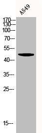
![Various whole cell extracts (30 μg) were separated by 7.5% SDS-PAGE, and the membrane was blotted with AKT antibody [N3C2], Internal (GTX121937) diluted at 1:1000. The HRP-conjugated anti-rabbit IgG antibody (GTX213110-01) was used to detect the primary antibody, and the signal was developed with Trident ECL plus-Enhanced.](https://www.genetex.com/upload/website/prouct_img/normal/GTX121937/GTX121937_43264_20180706_WB_M_w_23060519_816.webp "Various whole cell extracts (30 μg) were separated by 7.5% SDS-PAGE, and the membrane was blotted with AKT antibody [N3C2], Internal (GTX121937) diluted at 1:1000. The HRP-conjugated anti-rabbit IgG antibody (GTX213110-01) was used to detect the primary antibody, and the signal was developed with Trident ECL plus-Enhanced.")
![Immunoprecipitation of Akt1/2/3 protein from 293T whole cell extracts using 5 μg of Akt1/2/3 antibody [N3C2], Internal (GTX121937). Western blot analysis was performed using Akt1/2/3 antibody [N3C2], Internal (GTX121937). EasyBlot anti-Rabbit IgG (GTX221666-01) was used as a secondary reagent.](https://www.genetex.com/upload/website/prouct_img/normal/GTX121937/GTX121937_40450_20150327_IP_w_23060519_222.webp "Immunoprecipitation of Akt1/2/3 protein from 293T whole cell extracts using 5 μg of Akt1/2/3 antibody [N3C2], Internal (GTX121937). Western blot analysis was performed using Akt1/2/3 antibody [N3C2], Internal (GTX121937). EasyBlot anti-Rabbit IgG (GTX221666-01) was used as a secondary reagent.")
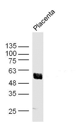
![Non-transfected (–) and transfected (+) 293T whole cell extracts (30 μg) were separated by 10% SDS-PAGE, and the membrane was blotted with AKT antibody [N3C2], Internal (GTX121937) diluted at 1:1000. The HRP-conjugated anti-rabbit IgG antibody (GTX213110-01) was used to detect the primary antibody, and the signal was developed with Trident ECL plus-Enhanced.](https://www.genetex.com/upload/website/prouct_img/normal/GTX121937/GTX121937_43264_20180824_WB_B_w_23060519_793.webp "Non-transfected (–) and transfected (+) 293T whole cell extracts (30 μg) were separated by 10% SDS-PAGE, and the membrane was blotted with AKT antibody [N3C2], Internal (GTX121937) diluted at 1:1000. The HRP-conjugated anti-rabbit IgG antibody (GTX213110-01) was used to detect the primary antibody, and the signal was developed with Trident ECL plus-Enhanced.")
 A: JC B: BCL-1 7.5% SDS PAGE GTX121937 diluted at 1:5000 The HRP-conjugated anti-rabbit IgG antibody (GTX213110-01) was used to detect the primary antibody.")


Various whole cell extracts (30 μg) were separated by 7.5% SDS-PAGE, and the membrane was blotted with AKT antibody [N3C2], Internal (GTX121937) diluted at 1:3000. The HRP-conjugated anti-rabbit IgG antibody (GTX213110-01) was used to detect the primary antibody, and the signal was developed with Trident ECL plus-Enhanced.
AKT antibody [N3C2], Internal
GTX121937
ApplicationsImmunoFluorescence, ImmunoPrecipitation, Western Blot, ImmunoCytoChemistry, ImmunoHistoChemistry, ImmunoHistoChemistry Frozen, ImmunoHistoChemistry Paraffin
Product group Antibodies
ReactivityFish, Human, Mouse, Porcine, Rat
TargetAKT1
Overview
- SupplierGeneTex
- Product NameAKT antibody [N3C2], Internal
- Delivery Days Customer9
- Application Supplier NoteWB: 1:500-1:3000. ICC/IF: 1:100-1:1000. IP: 1:100-1:500. *Optimal dilutions/concentrations should be determined by the researcher.Not tested in other applications.
- ApplicationsImmunoFluorescence, ImmunoPrecipitation, Western Blot, ImmunoCytoChemistry, ImmunoHistoChemistry, ImmunoHistoChemistry Frozen, ImmunoHistoChemistry Paraffin
- CertificationResearch Use Only
- ClonalityPolyclonal
- Concentration0.67 mg/ml
- ConjugateUnconjugated
- Gene ID207
- Target nameAKT1
- Target descriptionAKT serine/threonine kinase 1
- Target synonymsAKT, PKB, PKB-ALPHA, PRKBA, RAC, RAC-ALPHA, RAC-alpha serine/threonine-protein kinase, AKT1m, PKB alpha, RAC-PK-alpha, protein kinase B alpha, proto-oncogene c-Akt, rac protein kinase alpha, serine-threonine protein kinase, v-akt murine thymoma viral oncogene homolog 1, v-akt murine thymoma viral oncogene-like protein 1
- HostRabbit
- IsotypeIgG
- Scientific DescriptionThe serine-threonine protein kinase encoded by the AKT1 gene is catalytically inactive in serum-starved primary and immortalized fibroblasts. AKT1 and the related AKT2 are activated by platelet-derived growth factor. The activation is rapid and specific, and it is abrogated by mutations in the pleckstrin homology domain of AKT1. It was shown that the activation occurs through phosphatidylinositol 3-kinase. In the developing nervous system AKT is a critical mediator of growth factor-induced neuronal survival. Survival factors can suppress apoptosis in a transcription-independent manner by activating the serine/threonine kinase AKT1, which then phosphorylates and inactivates components of the apoptotic machinery. Mutations in this gene have been associated with the Proteus syndrome. Multiple alternatively spliced transcript variants have been found for this gene. [provided by RefSeq, Jul 2011]
- ReactivityFish, Human, Mouse, Porcine, Rat
- Storage Instruction-20°C or -80°C,2°C to 8°C
- UNSPSC41116161
Datasheet
Related products
Product group Antibodies
Anti-AKT1 AntibodyA285951
ApplicationsWestern Blot, ELISA
ReactivityHuman, Mouse
- SizePrice
Product group Antibodies
Anti-AKT1 Antibody144-11016
ApplicationsImmunoFluorescence, Western Blot, ImmunoHistoChemistry
ReactivityHuman, Mouse, Rat
TargetAKT1
- SizePrice
Product group Antibodies

Anti-pAKT [AbAb-pAKT]AB04102-10.0
ApplicationsImmunoPrecipitation, Western Blot, ELISA
ReactivityHuman, Mouse
TargetAKT1
- SizePrice

Product group Antibodies
Anti-AKT1 AntibodyAMAB90834
ApplicationsWestern Blot, ImmunoCytoChemistry
ReactivityHuman
TargetAKT1
- SizePrice
Product group Antibodies

AKT1 AntibodyLS-C831478
ApplicationsImmunoHistoChemistry
ReactivityHuman
TargetAKT1
- SizePrice

Product group Antibodies
Anti-AKT1,2,3 Antibody Picoband(r)A00024-2-CARRIER-FREE
ApplicationsFlow Cytometry, ImmunoFluorescence, Western Blot, ELISA, ImmunoCytoChemistry
ReactivityHuman, Mouse, Rat
TargetAKT1
- SizePrice
Product group Antibodies
AKT1 Polyclonal AntibodyBS-0115M
ApplicationsImmunoFluorescence, Western Blot, ELISA, ImmunoCytoChemistry, ImmunoHistoChemistry, ImmunoHistoChemistry Frozen, ImmunoHistoChemistry Paraffin
ReactivityBovine, Canine, Chicken, Human, Mouse, Porcine, Rabbit, Rat, Sheep
TargetAKT1
- SizePrice
Product group Antibodies
AKT1/AKT2/AKT3 AntibodyCSB-PA000848
ApplicationsWestern Blot, ELISA, ImmunoHistoChemistry
ReactivityHuman, Mouse, Rat
TargetAKT1
- SizePrice
Product group Antibodies

Goat anti-AKT1EB06875
ApplicationsWestern Blot, ELISA
ReactivityBovine, Human, Mouse
TargetAKT1
- SizePrice