

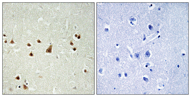
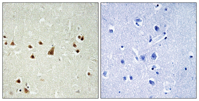
ANKRD26 antibody
GTX128255
ApplicationsImmunoFluorescence, Western Blot, ImmunoCytoChemistry
Product group Antibodies
TargetANKRD26
Overview
- SupplierGeneTex
- Product NameANKRD26 antibody
- Delivery Days Customer9
- Application Supplier NoteWB: 1:500-1:3000. *Optimal dilutions/concentrations should be determined by the researcher.Not tested in other applications.
- ApplicationsImmunoFluorescence, Western Blot, ImmunoCytoChemistry
- CertificationResearch Use Only
- ClonalityPolyclonal
- Concentration1 mg/ml
- ConjugateUnconjugated
- Gene ID22852
- Target nameANKRD26
- Target descriptionankyrin repeat domain containing 26
- Target synonymsTHC2, bA145E8.1, ankyrin repeat domain-containing protein 26, GNS/ANKRD26/NCKAP1L fusion, ankyrin repeat domain 26
- HostRabbit
- IsotypeIgG
- Protein IDQ9UPS8
- Protein NameAnkyrin repeat domain-containing protein 26
- Scientific DescriptionThis gene encodes a protein containing N-terminal ankyrin repeats which function in protein-protein interactions. Mutations in this gene are associated with autosomal dominant thrombocytopenia-2. Pseudogenes of this gene are found on chromosome 7, 10, 13 and 16. Multiple transcript variants encoding different isoforms have been found for this gene. [provided by RefSeq, Dec 2011]
- Storage Instruction-20°C or -80°C,2°C to 8°C
- UNSPSC12352203
References
- Carden S, Vitiello E, Rosa E Silva I, et al. Proteomic profiling of centrosomes across multiple mammalian cell and tissue types by an affinity capture method. Dev Cell. 2023,58(21):2393-2410.e9. doi: 10.1016/j.devcel.2023.09.008Read this paper
- Hall EA, Kumar D, Prosser SL, et al. Centriolar satellites expedite mother centriole remodeling to promote ciliogenesis. Elife. 2023,12. doi: 10.7554/eLife.79299Read this paper
- Evans LT, Anglen T, Scott P, et al. ANKRD26 recruits PIDD1 to centriolar distal appendages to activate the PIDDosome following centrosome amplification. EMBO J. 2021,40(4):e105106. doi: 10.15252/embj.2020105106Read this paper
- Yan H, Chen C, Chen H, et al. TALPID3 and ANKRD26 selectively orchestrate FBF1 localization and cilia gating. Nat Commun. 2020,11(1):2196. doi: 10.1038/s41467-020-16042-wRead this paper
- Sahabandu N, Kong D, Magidson V, et al. Expansion microscopy for the analysis of centrioles and cilia. J Microsc. 2019,276(3):145-159. doi: 10.1111/jmi.12841Read this paper
- Bowler M, Kong D, Sun S, et al. High-resolution characterization of centriole distal appendage morphology and dynamics by correlative STORM and electron microscopy. Nat Commun. 2019,10(1):993. doi: 10.1038/s41467-018-08216-4Read this paper
Datasheet
Related products
Product group Antibodies
ANKRD26 Polyclonal AntibodyBS-9756R
ApplicationsImmunoFluorescence, Western Blot, ImmunoHistoChemistry, ImmunoHistoChemistry Paraffin
TargetANKRD26
- SizePrice
Product group Antibodies
ANKRD26 antibodyGTX87200
ApplicationsImmunoHistoChemistry, ImmunoHistoChemistry Paraffin
TargetANKRD26
- SizePrice
Product group Antibodies

ANKRD26 Antibody (aa1659-1709)LS-C289058
ApplicationsImmunoPrecipitation, Western Blot
TargetANKRD26
- SizePrice

Product group Antibodies
Anti-ANKRD26 AntibodyHPA040654
ApplicationsImmunoCytoChemistry, ImmunoHistoChemistry
ReactivityHuman
TargetANKRD26
- SizePrice
Product group Antibodies
ANKRD26 AntibodyCSB-PA008262
ApplicationsELISA, ImmunoHistoChemistry
ReactivityHuman
TargetANKRD26
- SizePrice

Product group Antibodies
Anti-ANKRD26 AntibodyA09289-1
ApplicationsImmunoFluorescence, ELISA, ImmunoHistoChemistry
TargetANKRD26
- SizePrice