

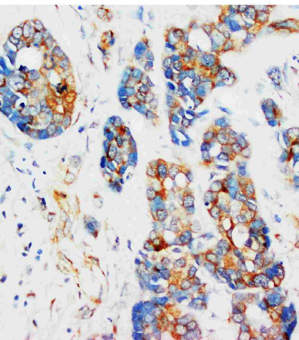
Figure 1. IHC analysis of alpha-Tubulin using anti-alpha-Tubulin antibody (MA1107). alpha-Tubulin was detected in a paraffin-embedded section of human mammary cancer tissue. Heat mediated antigen retrieval was performed in EDTA buffer (pH 8.0, epitope retrieval solution). The tissue section was blocked with 10% goat serum. The tissue section was then incubated with 1 microg/ml mouse anti-alpha-Tubulin Antibody (MA1107) overnight at 4°C. Peroxidase Conjugated Goat Anti-mouse IgG was used as secondary antibody and incubated for 30 minutes at 37°C. The tissue section was developed using HRP Conjugated Mouse IgG Super Vision Assay Kit (Catalog # SV0001) with DAB as the chromogen.
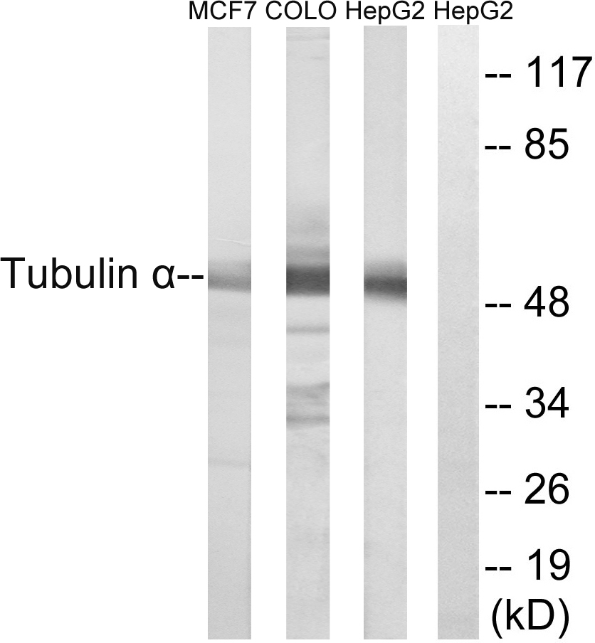
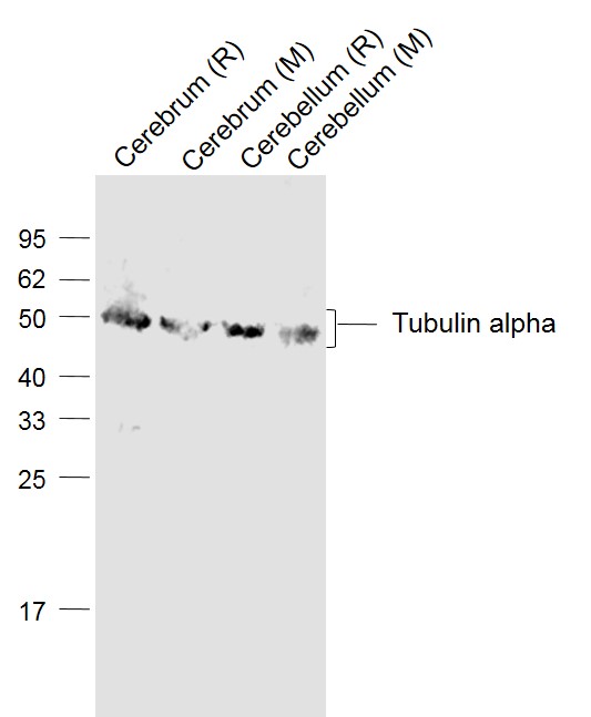
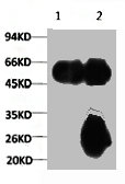
. Electrophoresis was performed on a 5-20% SDS-PAGE gel at 70V (Stacking gel) / 90V (Resolving gel) for 2-3 hours. The sample well of each lane was loaded with 30 ug of sample under reducing conditions. Lane 1: human SH-SY5Y whole cell lysates, Lane 2: human Jurkat whole cell lysates, Lane 3: human A549 whole cell lysates, Lane 4: rat brain tissue lysates, Lane 5: mouse brain tissue lysates, Lane 6: mouse thymus tissue lysates. After electrophoresis, proteins were transferred to a nitrocellulose membrane at 150 mA for 50-90 minutes. Blocked the membrane with 5% non-fat milk/TBS for 1.5 hour at RT. The membrane was incubated with mouse anti-alpha-Tubulin antigen affinity purified monoclonal antibody (Catalog # MA1107) at 1 microg/mL overnight at 4°C, then washed with TBS-0.1%Tween 3 times with 5 minutes each and probed with a goat anti-mouse IgG-HRP secondary antibody at a dilution of 1:10000 for 1.5 hour at RT. The signal is developed using an Enhanced Chemiluminescent detection (ECL) kit (Catalog # EK1001) with Tanon 5200 system. A specific band was detected for alpha-Tubulin at approximately 55 kDa. The expected band size for alpha-Tubulin is at 50 kDa.")
Figure 1. IHC analysis of alpha-Tubulin using anti-alpha-Tubulin antibody (MA1107). alpha-Tubulin was detected in a paraffin-embedded section of human mammary cancer tissue. Heat mediated antigen retrieval was performed in EDTA buffer (pH 8.0, epitope retrieval solution). The tissue section was blocked with 10% goat serum. The tissue section was then incubated with 1 microg/ml mouse anti-alpha-Tubulin Antibody (MA1107) overnight at 4°C. Peroxidase Conjugated Goat Anti-mouse IgG was used as secondary antibody and incubated for 30 minutes at 37°C. The tissue section was developed using HRP Conjugated Mouse IgG Super Vision Assay Kit (Catalog # SV0001) with DAB as the chromogen.
Anti-Alpha-Tubulin TUBA1A Antibody (Monoclonal, DM1A)
MA1107
ApplicationsWestern Blot, ImmunoHistoChemistry
Product group Antibodies
ReactivityChicken, Human, Mouse, Rat
TargetTUBA1A
Overview
- SupplierBoster Bio
- Product NameAnti-Alpha-Tubulin TUBA1A Antibody (Monoclonal, DM1A)
- Delivery Days Customer9
- Application Supplier NoteOther applications have not been tested. Optimal dilutions should be determined by end users.
- ApplicationsWestern Blot, ImmunoHistoChemistry
- Applications SupplierIHP, WB, IHC
- CertificationResearch Use Only
- ClonalityMonoclonal
- Clone IDDM1A
- Concentration100 ug/ml
- Gene ID7846
- Target nameTUBA1A
- Target descriptiontubulin alpha 1a
- Target synonymsB-ALPHA-1, LIS3, TUBA3, tubulin alpha-1A chain, hum-a-tub1, hum-a-tub2, tubulin B-alpha-1, tubulin alpha-3 chain, tubulin, alpha, brain-specific
- HostMouse
- IsotypeIgG1
- Protein IDQ71U36
- Protein NameTubulin alpha-1A chain
- Scientific DescriptionBoster Bio Anti-Alpha-Tubulin TUBA1A Antibody (Monoclonal, DM1A) catalog # MA1107. Tested in IHC, WB applications. This antibody reacts with Chicken, Human, Mouse, Rat.
- ReactivityChicken, Human, Mouse, Rat
- Reactivity SupplierChicken, Human, Mouse, Rat, Chicken
- Storage Instruction-20°C,2°C to 8°C
- UNSPSC12352203
References
- Wang F, Zhang S, Sun F, et al. Anti-angiogenesis and anti-immunosuppression gene therapy through targeting COUP-TFII in an in situ glioblastoma mouse model. Cancer Gene Ther. 2024,31(8):1135-1150. doi: 10.1038/s41417-024-00799-zRead this paper
- Li X, Yuan F, Xiong Y, et al. FAM3A plays a key role in protecting against tubular cell pyroptosis and acute kidney injury. Redox Biol. 2024,74:103225. doi: 10.1016/j.redox.2024.103225Read this paper
- Huang Q, Liu L, Tan X, et al. Empagliflozin alleviates neuroinflammation by inhibiting astrocyte activation in the brain and regulating gut microbiota of high-fat diet mice. J Affect Disord. 2024,360:229-241. doi: 10.1016/j.jad.2024.05.150Read this paper
- Song X, Li D, Gan L, et al. Intravenous Injection of Na Ions Aggravates Ang II-Induced Hypertension-Related Vascular Endothelial Injury by Increasing Transmembrane Osmotic Pressure. Int J Nanomedicine. 2023,18:7505-7521. doi: 10.2147/IJN.S435144Read this paper
- Li H, Zhu L, Weng Z, et al. Sesamin attenuates UVA-induced keratinocyte injury via inhibiting ASK-1-JNK/p38 MAPK pathways. J Cosmet Dermatol. 2024,23(1):316-325. doi: 10.1111/jocd.15951Read this paper
- Song J, Liu S, Ren Y, et al. Organotin benzohydroxamate derivatives (OTBH) target colchicine-binding site exerting potent antitumor activity both in vitro and vivo revealed by quantitative proteomic analysis. Eur J Pharm Sci. 2023,187:106488. doi: 10.1016/j.ejps.2023.106488Read this paper
- Dong H, Zeng L, Chen W, et al. N6-methyladenine-mediated aberrant activation of the lncRNA SOX2OT-GLI1 loop promotes non-small-cell lung cancer stemness. Cell Death Discov. 2023,9(1):149. doi: 10.1038/s41420-023-01442-wRead this paper
- Hu Y, Xie Q, Wu X, et al. Tension of plus-end tracking protein Clip170 confers directionality and aggressiveness during breast cancer migration. Cell Death Dis. 2022,13(10):856. doi: 10.1038/s41419-022-05306-6Read this paper
- Zhang J, Hao N, Li W, et al. Simvastatin Upregulates Lipoxin A4 and Accelerates Neuroinflammation Resolution After Intracerebral Hemorrhage. Curr Neurovasc Res. 2022,19(3):321-332. doi: 10.2174/1567202619666220913124627Read this paper
- Huang J, Xu Z, Chen H, et al. Shen Qi Wan Ameliorates Learning and Memory Impairment Induced by STZ in AD Rats through PI3K/AKT Pathway. Brain Sci. 2022,12(6). doi: 10.3390/brainsci12060758Read this paper
Datasheet
MSDS
Related products
Product group Antibodies
ApplicationsImmunoFluorescence, Western Blot, ELISA, ImmunoHistoChemistry
ReactivityHuman, Mouse, Rat
- SizePrice
Product group Antibodies

Anti-Alpha-Tubulin [F2C]Ab00403-1.1
ApplicationsImmunoFluorescence, Western Blot, ELISA
ReactivityHuman
TargetTUBA1A
- SizePrice
Product group Antibodies
Anti-Tubulin alpha Antibody102-25843
ApplicationsImmunoFluorescence, Western Blot, ImmunoHistoChemistry
TargetTUBA1A
- SizePrice

Product group Antibodies
Anti-Tubulin alpha Antibody Picoband(r)A03989-1-CARRIER-FREE
ApplicationsFlow Cytometry, ImmunoFluorescence, Western Blot, ELISA, ImmunoCytoChemistry, ImmunoHistoChemistry
ReactivityHuman, Mouse, Rat
TargetTUBA1A
- SizePrice
Product group Antibodies
Tubulin alpha Polyclonal AntibodyBS-20496R
ApplicationsImmunoFluorescence, Western Blot, ELISA, ImmunoCytoChemistry, ImmunoHistoChemistry, ImmunoHistoChemistry Frozen, ImmunoHistoChemistry Paraffin
ReactivityHuman, Mouse, Rat
TargetTUBA1A
- SizePrice
Product group Antibodies
TUBA1A Monoclonal AntibodyCSB-MA000190
ApplicationsImmunoPrecipitation, Western Blot, ELISA
ReactivityHuman, Mouse, Rat
TargetTUBA1A
- SizePrice
Product group Antibodies
TUBA1A Monoclonal AntibodyCAC12980
ApplicationsFlow Cytometry, ImmunoFluorescence, ImmunoPrecipitation, Western Blot, ELISA, ImmunoHistoChemistry
ReactivityMouse, Rabbit, Rat
- SizePrice
Product group Antibodies

TUBA1A / Tubulin Alpha 1a Antibody (HRP)LS-C377875
ApplicationsELISA, ImmunoHistoChemistry
ReactivityHuman
TargetTUBA1A
- SizePrice

Product group Antibodies
Anti-TUBA1A AntibodyHPA039247
ApplicationsWestern Blot, ImmunoCytoChemistry, ImmunoHistoChemistry
ReactivityHuman, Mouse, Rat
TargetTUBA1A
- SizePrice
Product group Antibodies
alpha Tubulin 1A antibodyGTX109832
ApplicationsImmunoFluorescence, Western Blot, ImmunoCytoChemistry, ImmunoHistoChemistry, ImmunoHistoChemistry Paraffin
ReactivityDrosophila, Human, Mouse, Rat
TargetTUBA1A
- SizePrice