

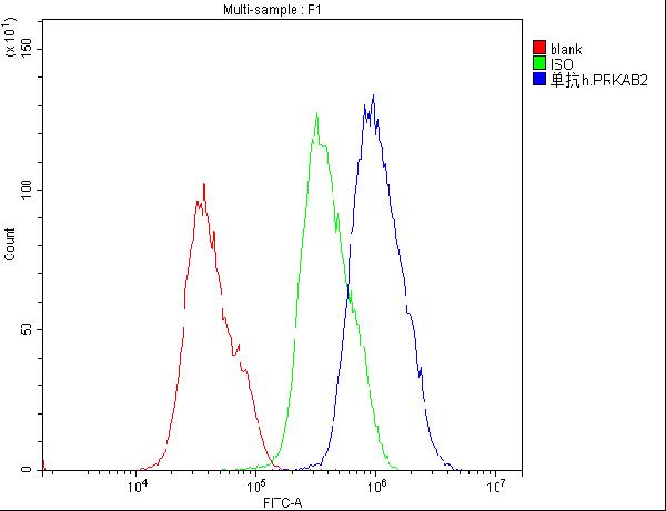
Figure 1. Flow Cytometry analysis of PC-3 cells using anti-AMPK beta 2 antibody (M05077). Overlay histogram showing PC-3 cells stained with M05077 (Blue line). To facilitate intracellular staining, cells were fixed with 4% paraformaldehyde and permeabilized with permeabilization buffer. The cells were blocked with 10% normal goat serum. And then incubated with mouse anti-AMPK beta 2 Antibody (M05077,1microg/1x106 cells) for 30 min at 20°C. DyLight®488 conjugated goat anti-mouse IgG (BA1126, 5-10microg/1x106 cells) was used as secondary antibody for 30 minutes at 20°C. Isotype control antibody (Green line) was mouse IgG (1microg/1x106) used under the same conditions. Unlabelled sample (Red line) was also used as a control.
. Electrophoresis was performed on a 10% SDS-PAGE gel at 70V (Stacking gel) / 90V (Resolving gel) for 2-3 hours. The sample well of each lane was loaded with 50ug of sample under reducing conditions. Lane 1: human Hela whole cell lysate, Lane 2: human placenta tissue lysate, Lane 3: human 293T whole cell lysate, Lane 4: human A549 whole cell lysate, Lane 5: human A375 whole cell lysate, Lane 6: human A431 whole cell lysate, Lane 7: human U20S whole cell lysate, Lane 8: human K562 whole cell lysate. After Electrophoresis, proteins were transferred to a Nitrocellulose membrane at 150mA for 50-90 minutes. Blocked the membrane with 5% Non-fat Milk/ TBS for 1.5 hour at RT. The membrane was incubated with mouse anti-AMPK beta 2 antigen affinity purified monoclonal antibody (Catalog # M05077) at 0.5 microg/mL overnight at 4°C, then washed with TBS-0.1%Tween 3 times with 5 minutes each and probed with a goat anti-mouse IgG-HRP secondary antibody at a dilution of 1:10000 for 1.5 hour at RT. The signal is developed using an Enhanced Chemiluminescent detection (ECL) kit (Catalog # EK1001) with Tanon 5200 system.")
Figure 1. Flow Cytometry analysis of PC-3 cells using anti-AMPK beta 2 antibody (M05077). Overlay histogram showing PC-3 cells stained with M05077 (Blue line). To facilitate intracellular staining, cells were fixed with 4% paraformaldehyde and permeabilized with permeabilization buffer. The cells were blocked with 10% normal goat serum. And then incubated with mouse anti-AMPK beta 2 Antibody (M05077,1microg/1x106 cells) for 30 min at 20°C. DyLight®488 conjugated goat anti-mouse IgG (BA1126, 5-10microg/1x106 cells) was used as secondary antibody for 30 minutes at 20°C. Isotype control antibody (Green line) was mouse IgG (1microg/1x106) used under the same conditions. Unlabelled sample (Red line) was also used as a control.
Anti-AMPK beta 2 PRKAB2 Antibody Picoband(r) (monoclonal, 6G1)
M05077-10UG
ApplicationsFlow Cytometry, Western Blot, ImmunoCytoChemistry, ImmunoHistoChemistry
Product group Antibodies
ReactivityHuman
TargetPRKAB2
Overview
- SupplierBoster Bio
- Product NameAnti-AMPK beta 2 PRKAB2 Antibody Picoband(r) (monoclonal, 6G1)
- Delivery Days Customer9
- Antibody SpecificityNo cross reactivity with other proteins.
- ApplicationsFlow Cytometry, Western Blot, ImmunoCytoChemistry, ImmunoHistoChemistry
- CertificationResearch Use Only
- ClonalityMonoclonal
- Clone ID6G1
- Concentration500 ug/ml
- Gene ID5565
- Target namePRKAB2
- Target descriptionprotein kinase AMP-activated non-catalytic subunit beta 2
- Target synonyms5'-AMP-activated protein kinase subunit beta-2; 5'-AMP-activated protein kinase, beta-2 subunit; AMP-activated protein kinase beta 2 non-catalytic subunit; AMPK beta 2; AMPK beta-2 chain; AMPK subunit beta-2; protein kinase, AMP-activated, beta 2 non-catalytic subunit
- HostMouse
- IsotypeIgG2b
- Protein IDO43741
- Protein Name5'-AMP-activated protein kinase subunit beta-2
- Scientific DescriptionBoster Bio Anti-AMPK beta 2 PRKAB2 Antibody Picoband® (monoclonal, 6G1) catalog # M05077. Tested in Flow Cytometry, IHC, ICC, WB applications. This antibody reacts with Human. The brand Picoband indicates this is a premium antibody that guarantees superior quality, high affinity, and strong signals with minimal background in Western blot applications. Only our best-performing antibodies are designated as Picoband, ensuring unmatched performance.
- ReactivityHuman
- Storage Instruction-20°C,2°C to 8°C
- UNSPSC12352203