

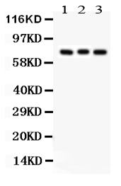
Figure 1. Western blot analysis of APG7 using anti-APG7 antibody (PB9479). Electrophoresis was performed on a 5-20% SDS-PAGE gel at 70V (Stacking gel) / 90V (Resolving gel) for 2-3 hours. Lane 1: Rat Brain Tissue Lysate at 50ug, Lane 2: Mouse Brain Tissue Lysate at 50ug, Lane 3: 293T Whole Cell Lysate at 40ug. After electrophoresis, proteins were transferred to a nitrocellulose membrane at 150 mA for 50-90 minutes. Blocked the membrane with 5% non-fat milk/TBS for 1.5 hour at RT. The membrane was incubated with rabbit anti-APG7 antigen affinity purified polyclonal antibody (Catalog # PB9479) at 0.5 microg/mL overnight at 4°C, then washed with TBS-0.1%Tween 3 times with 5 minutes each and probed with a goat anti-rabbit IgG-HRP secondary antibody at a dilution of 1:5000 for 1.5 hour at RT. The signal is developed using an Enhanced Chemiluminescent detection (ECL) kit (Catalog # EK1002) with Tanon 5200 system. A specific band was detected for APG7 at approximately 78 kDa. The expected band size for APG7 is at 78 kDa.
Figure 1. Western blot analysis of APG7 using anti-APG7 antibody (PB9479). Electrophoresis was performed on a 5-20% SDS-PAGE gel at 70V (Stacking gel) / 90V (Resolving gel) for 2-3 hours. Lane 1: Rat Brain Tissue Lysate at 50ug, Lane 2: Mouse Brain Tissue Lysate at 50ug, Lane 3: 293T Whole Cell Lysate at 40ug. After electrophoresis, proteins were transferred to a nitrocellulose membrane at 150 mA for 50-90 minutes. Blocked the membrane with 5% non-fat milk/TBS for 1.5 hour at RT. The membrane was incubated with rabbit anti-APG7 antigen affinity purified polyclonal antibody (Catalog # PB9479) at 0.5 microg/mL overnight at 4°C, then washed with TBS-0.1%Tween 3 times with 5 minutes each and probed with a goat anti-rabbit IgG-HRP secondary antibody at a dilution of 1:5000 for 1.5 hour at RT. The signal is developed using an Enhanced Chemiluminescent detection (ECL) kit (Catalog # EK1002) with Tanon 5200 system. A specific band was detected for APG7 at approximately 78 kDa. The expected band size for APG7 is at 78 kDa.
Anti-Apg7 Picoband Antibody
PB9479
ApplicationsWestern Blot
Product group Antibodies
ReactivityChicken, Human, Mouse, Rat
TargetATG7
Overview
- SupplierBoster Bio
- Product NameAnti-Apg7 Picoband Antibody
- Delivery Days Customer9
- Application Supplier NoteTested Species: In-house tested species with positive results. Other applications have not been tested. Optimal dilutions should be determined by end users.
- ApplicationsWestern Blot
- CertificationResearch Use Only
- ClonalityPolyclonal
- Concentration500 ug/ml
- Gene ID10533
- Target nameATG7
- Target descriptionautophagy related 7
- Target synonymsAPG7-LIKE, APG7L, GSA7, SCAR31, ubiquitin-like modifier-activating enzyme ATG7, APG7 autophagy 7-like, ATG12-activating enzyme E1 ATG7, hAGP7, ubiquitin-activating enzyme E1-like protein
- HostRabbit
- IsotypeIgG
- Protein IDO95352
- Protein NameUbiquitin-like modifier-activating enzyme ATG7
- Scientific DescriptionBoster Bio Anti-Apg7/ATG7 Antibody Picoband® catalog # PB9479. Tested in WB applications. This antibody reacts with Human, Mouse, Rat. The brand Picoband indicates this is a premium antibody that guarantees superior quality, high affinity, and strong signals with minimal background in Western blot applications. Only our best-performing antibodies are designated as Picoband, ensuring unmatched performance.
- ReactivityChicken, Human, Mouse, Rat
- Storage Instruction-20°C,2°C to 8°C
- UNSPSC12352203
References
- Shi Q, Li Z, Dong Y, et al. LncRNA THRIL, transcriptionally activated by AP-1 and stabilized by METTL14-mediated m6A modification, accelerates LPS-evoked acute injury in alveolar epithelial cells. Int Immunopharmacol. 2023,123:110740. doi: 10.1016/j.intimp.2023.110740Read this paper
- Yi J, Wang X, Song K, et al. Integrated metabolomics and mechanism to reveal the protective effect of kaempferol on pulmonary arterial hypertension. J Pharm Biomed Anal. 2022,212:114662. doi: 10.1016/j.jpba.2022.114662Read this paper
- Ren HH, Niu Z, Guo R, et al. Rhodiola crenulata extract decreases fatty acid oxidation and autophagy to ameliorate pulmonary arterial hypertension by targeting inhibiton of acylcarnitine in rats. Chin J Nat Med. 2021,19(2):120-133. doi: 10.1016/S1875-5364(21)60013-4Read this paper
- Zhong G , Yang X , Jiang X , et al. Dopamine-melanin nanoparticles scavenge reactive oxygen and nitrogen species and activate autophagy for osteoarthritis therapy. Nanoscale. 2019,11(24):11605-11616. doi: 10.1039/c9nr03060cRead this paper
- Xing JJ, Hou JG, Ma ZN, et al. Ginsenoside Rb3 provides protective effects against cisplatin-induced nephrotoxicity via regulation of AMPK-/mTOR-mediated autophagy and inhibition of apoptosis in vitro and in vivo. Cell Prolif. 2019,52(4):e12627. doi: 10.1111/cpr.12627Read this paper
- Zhang Y, Liu Y, Zou J, et al. Tetrahydrocurcumin induces mesenchymal-epithelial transition and suppresses angiogenesis by targeting HIF-1α and autophagy in human osteosarcoma. Oncotarget. 2017,8(53):91134-91149. doi: 10.18632/oncotarget.19845Read this paper
- Yang J, Sheng S, Yang Q, et al. Endocan silencing induces programmed cell death in hepatocarcinoma. Oncol Lett. 2017,14(5):5333-5339. doi: 10.3892/ol.2017.6857Read this paper
- You T, Cheng Y, Zhong J, et al. Roflupram, a Phosphodiesterase 4 Inhibitior, Suppresses Inflammasome Activation through Autophagy in Microglial Cells. ACS Chem Neurosci. 2017,8(11):2381-2392. doi: 10.1021/acschemneuro.7b00065Read this paper
Datasheet
MSDS
Related products

Product group Antibodies
Anti-ATG7 Antibody Picoband(r)A00346-4-CARRIER-FREE
ApplicationsFlow Cytometry, Western Blot, ELISA
ReactivityHuman
TargetATG7
- SizePrice
Product group Antibodies
Anti-ATG7 Antibody144-00691
ApplicationsWestern Blot, ImmunoHistoChemistry
ReactivityHuman, Mouse, Rat
TargetATG7
- SizePrice
Product group Antibodies
Atg7 Polyclonal AntibodyCAC08251
ApplicationsImmunoFluorescence, Western Blot, ELISA, ImmunoHistoChemistry
TargetATG7
- SizePrice
Product group Antibodies
References
ATG7/APG7 Polyclonal AntibodyBS-2432R
ApplicationsImmunoFluorescence, Western Blot, ELISA, ImmunoCytoChemistry, ImmunoHistoChemistry, ImmunoHistoChemistry Frozen, ImmunoHistoChemistry Paraffin
ReactivityBovine, Canine, Chicken, Equine, Human, Mouse, Porcine, Rat
TargetATG7
- SizePrice
Product group Antibodies
ATG7 Monoclonal AntibodyCSB-MA617582
ApplicationsELISA, ImmunoHistoChemistry
ReactivityHuman, Mouse, Rat
TargetATG7
- SizePrice
Product group Antibodies

Apg7 / ATG7 Antibody (clone 1A1)LS-C764703
ApplicationsImmunoFluorescence, ImmunoHistoChemistry, ImmunoHistoChemistry Paraffin
ReactivityHuman, Mouse, Rat
TargetATG7
- SizePrice

Product group Antibodies
Anti-ATG7 AntibodyHPA007639
ApplicationsImmunoCytoChemistry, ImmunoHistoChemistry
ReactivityHuman
TargetATG7
- SizePrice
![Whole cell extract (30 μg) was separated by 7.5% SDS-PAGE, and the membrane was blotted with ATG7 antibody [N3C2], Internal (GTX113613) diluted at 1:1000. The HRP-conjugated anti-rabbit IgG antibody (GTX213110-01) was used to detect the primary antibody, and the signal was developed with Trident ECL plus-Enhanced.](https://www.genetex.com/upload/website/prouct_img/normal/GTX113613/GTX113613_40142_20190913_WB_M_w_23060501_290.webp)
Product group Antibodies
References
ATG7 antibody [N3C2], InternalGTX113613
ApplicationsWestern Blot
ReactivityHuman, Mouse
TargetATG7
- SizePrice