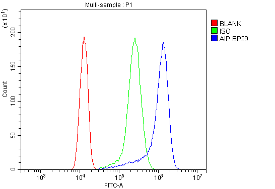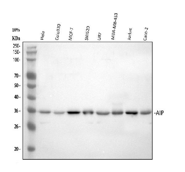
Figure 2. IF analysis of ARA9 using anti-ARA9 antibody (PB9042). ARA9 was detected in immunocytochemical section of U20S cells. Enzyme antigen retrieval was performed using IHC enzyme antigen retrieval reagent (AR0022) for 15 mins. The cells were blocked with 10% goat serum. And then incubated with 2microg/mL rabbit anti-ARA9 Antibody (PB9042) overnight at 4°C. DyLight®488 Conjugated Goat Anti-Rabbit IgG (BA1127) was used as secondary antibody at 1:100 dilution and incubated for 30 minutes at 37°C. The section was counterstained with DAPI. Visualize using a fluorescence microscope and filter sets appropriate for the label used.
Anti-ARA9/AIP Antibody Picoband(r)

PB9042
ApplicationsFlow Cytometry, ImmunoFluorescence, Western Blot, ImmunoCytoChemistry
Product group Antibodies
ReactivityHuman
TargetAIP
Overview
- SupplierBoster Bio
- Product NameAnti-ARA9 Picoband Antibody
- Delivery Days Customer9
- Antibody SpecificityNo cross reactivity with other proteins.
- Application Supplier NoteWB: The detection limit for ARA9 is approximately 0.25ng/lane under reducing conditions. Tested Species: In-house tested species with positive results. Other applications have not been tested. Optimal dilutions should be determined by end users.
- ApplicationsFlow Cytometry, ImmunoFluorescence, Western Blot, ImmunoCytoChemistry
- Applications SupplierWB
- CertificationResearch Use Only
- ClonalityPolyclonal
- Concentration500 ug/ml
- FormulationLyophilized
- Gene ID9049
- Target nameAIP
- Target descriptionaryl hydrocarbon receptor interacting protein
- Target synonymsAh receptor activated 9; AH receptor-interacting protein; ARA9; aryl hydrocarbon receptor-associated protein 9; FK506-binding protein 37; FKBP prolyl isomerase 16; FKBP16; FKBP37; HBV X-associated protein 2; hepatitis B virus X-associated cellular protein 2; immunophilin homolog ARA9; PITA1; SMTPHN; XAP2; XAP-2; X-associated protein-2
- HostRabbit
- IsotypeIgG
- Protein IDO00170
- Protein NameAH receptor-interacting protein
- Scientific DescriptionBoster Bio Anti-ARA9/AIP Antibody Picoband® catalog # PB9042. Tested in Flow Cytometry, IF, ICC, WB applications. This antibody reacts with Human. The brand Picoband indicates this is a premium antibody that guarantees superior quality, high affinity, and strong signals with minimal background in Western blot applications. Only our best-performing antibodies are designated as Picoband, ensuring unmatched performance.
- ReactivityHuman
- Reactivity SupplierHuman
- Storage Instruction-20°C,2°C to 8°C
- UNSPSC12352203


