

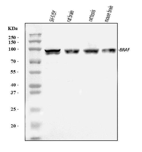
Figure 1. Western blot analysis of B Raf/BRAF using anti-B Raf/BRAF antibody (PB9164). Electrophoresis was performed on a 5-20% SDS-PAGE gel at 70V (Stacking gel) / 90V (Resolving gel) for 2-3 hours. The sample well of each lane was loaded with 30 ug of sample under reducing conditions. Lane 1: human SH-SY5Y whole cell lysates, Lane 2: rat brain tissue lysates, Lane 3: rat testis tissue lysates, Lane 4: mouse brain tissue lysates. After electrophoresis, proteins were transferred to a nitrocellulose membrane at 150 mA for 50-90 minutes. Blocked the membrane with 5% non-fat milk/TBS for 1.5 hour at RT. The membrane was incubated with rabbit anti-B Raf/BRAF antigen affinity purified polyclonal antibody (Catalog # PB9164) at 0.5 microg/mL overnight at 4°C, then washed with TBS-0.1%Tween 3 times with 5 minutes each and probed with a goat anti-rabbit IgG-HRP secondary antibody at a dilution of 1:5000 for 1.5 hour at RT. The signal is developed using an Enhanced Chemiluminescent detection (ECL) kit (Catalog # EK1002) with Tanon 5200 system. A specific band was detected for B Raf/BRAF at approximately 84 kDa. The expected band size for B Raf/BRAF is at 84 kDa.
. B Raf/BRAF was detected in a paraffin-embedded section of human liver cancer tissue. Heat mediated antigen retrieval was performed in EDTA buffer (pH 8.0, epitope retrieval solution). The tissue section was blocked with 10% goat serum. The tissue section was then incubated with 2 microg/ml rabbit anti-B Raf/BRAF Antibody (PB9164) overnight at 4°C. Peroxidase Conjugated Goat Anti-rabbit IgG was used as secondary antibody and incubated for 30 minutes at 37°C. The tissue section was developed using HRP Conjugated Rabbit IgG Super Vision Assay Kit (Catalog # SV0002) with DAB as the chromogen.")
. B Raf/BRAF was detected in a paraffin-embedded section of human lung cancer tissue. Heat mediated antigen retrieval was performed in EDTA buffer (pH 8.0, epitope retrieval solution). The tissue section was blocked with 10% goat serum. The tissue section was then incubated with 2 microg/ml rabbit anti-B Raf/BRAF Antibody (PB9164) overnight at 4°C. Peroxidase Conjugated Goat Anti-rabbit IgG was used as secondary antibody and incubated for 30 minutes at 37°C. The tissue section was developed using HRP Conjugated Rabbit IgG Super Vision Assay Kit (Catalog # SV0002) with DAB as the chromogen.")
. B Raf/BRAF was detected in a paraffin-embedded section of human placenta tissue. Heat mediated antigen retrieval was performed in EDTA buffer (pH 8.0, epitope retrieval solution). The tissue section was blocked with 10% goat serum. The tissue section was then incubated with 2 microg/ml rabbit anti-B Raf/BRAF Antibody (PB9164) overnight at 4°C. Peroxidase Conjugated Goat Anti-rabbit IgG was used as secondary antibody and incubated for 30 minutes at 37°C. The tissue section was developed using HRP Conjugated Rabbit IgG Super Vision Assay Kit (Catalog # SV0002) with DAB as the chromogen.")
. B Raf/BRAF was detected in a paraffin-embedded section of human differentiated adenocarcinoma of the rectum tissue. Heat mediated antigen retrieval was performed in EDTA buffer (pH 8.0, epitope retrieval solution). The tissue section was blocked with 10% goat serum. The tissue section was then incubated with 2 microg/ml rabbit anti-B Raf/BRAF Antibody (PB9164) overnight at 4°C. Peroxidase Conjugated Goat Anti-rabbit IgG was used as secondary antibody and incubated for 30 minutes at 37°C. The tissue section was developed using HRP Conjugated Rabbit IgG Super Vision Assay Kit (Catalog # SV0002) with DAB as the chromogen.")
. B Raf/BRAF was detected in a paraffin-embedded section of mouse brain tissue. Heat mediated antigen retrieval was performed in EDTA buffer (pH 8.0, epitope retrieval solution). The tissue section was blocked with 10% goat serum. The tissue section was then incubated with 2 microg/ml rabbit anti-B Raf/BRAF Antibody (PB9164) overnight at 4°C. Peroxidase Conjugated Goat Anti-rabbit IgG was used as secondary antibody and incubated for 30 minutes at 37°C. The tissue section was developed using HRP Conjugated Rabbit IgG Super Vision Assay Kit (Catalog # SV0002) with DAB as the chromogen.")
. B Raf/BRAF was detected in a paraffin-embedded section of rat brain tissue. Heat mediated antigen retrieval was performed in EDTA buffer (pH 8.0, epitope retrieval solution). The tissue section was blocked with 10% goat serum. The tissue section was then incubated with 2 microg/ml rabbit anti-B Raf/BRAF Antibody (PB9164) overnight at 4°C. Peroxidase Conjugated Goat Anti-rabbit IgG was used as secondary antibody and incubated for 30 minutes at 37°C. The tissue section was developed using HRP Conjugated Rabbit IgG Super Vision Assay Kit (Catalog # SV0002) with DAB as the chromogen.")
. Overlay histogram showing U20S cells stained with PB9164 (Blue line). To facilitate intracellular staining, cells were fixed with 4% paraformaldehyde and permeabilized with permeabilization buffer. The cells were blocked with 10% normal goat serum. And then incubated with rabbit anti-B Raf/BRAF Antibody (PB9164, 1 microg/1x106 cells) for 30 min at 20°C. DyLight®488 conjugated goat anti-rabbit IgG (BA1127, 5-10 microg/1x106 cells) was used as secondary antibody for 30 minutes at 20°C. Isotype control antibody (Green line) was rabbit IgG (1 microg/1x106) used under the same conditions. Unlabelled sample without incubation with primary antibody and secondary antibody (Red line) was used as a blank control.")
Figure 1. Western blot analysis of B Raf/BRAF using anti-B Raf/BRAF antibody (PB9164). Electrophoresis was performed on a 5-20% SDS-PAGE gel at 70V (Stacking gel) / 90V (Resolving gel) for 2-3 hours. The sample well of each lane was loaded with 30 ug of sample under reducing conditions. Lane 1: human SH-SY5Y whole cell lysates, Lane 2: rat brain tissue lysates, Lane 3: rat testis tissue lysates, Lane 4: mouse brain tissue lysates. After electrophoresis, proteins were transferred to a nitrocellulose membrane at 150 mA for 50-90 minutes. Blocked the membrane with 5% non-fat milk/TBS for 1.5 hour at RT. The membrane was incubated with rabbit anti-B Raf/BRAF antigen affinity purified polyclonal antibody (Catalog # PB9164) at 0.5 microg/mL overnight at 4°C, then washed with TBS-0.1%Tween 3 times with 5 minutes each and probed with a goat anti-rabbit IgG-HRP secondary antibody at a dilution of 1:5000 for 1.5 hour at RT. The signal is developed using an Enhanced Chemiluminescent detection (ECL) kit (Catalog # EK1002) with Tanon 5200 system. A specific band was detected for B Raf/BRAF at approximately 84 kDa. The expected band size for B Raf/BRAF is at 84 kDa.
Anti-B Raf/BRAF Antibody Picoband(r)

PB9164
ApplicationsFlow Cytometry, Western Blot, ImmunoHistoChemistry
Product group Antibodies
ReactivityHuman, Mouse, Rat
TargetBRAF
Overview
- SupplierBoster Bio
- Product NameAnti-B Raf Picoband Antibody
- Delivery Days Customer9
- Antibody SpecificityNo cross reactivity with other proteins.
- Application Supplier NoteWB: The detection limit for B Raf is approximately 0.25ng/lane under reducing conditions. Tested Species: In-house tested species with positive results. Other applications have not been tested. Optimal dilutions should be determined by end users.
- ApplicationsFlow Cytometry, Western Blot, ImmunoHistoChemistry
- Applications SupplierWB
- CertificationResearch Use Only
- ClonalityPolyclonal
- Concentration500 ug/ml
- FormulationLyophilized
- Gene ID673
- Target nameBRAF
- Target descriptionB-Raf proto-oncogene, serine/threonine kinase
- Target synonyms94 kDa B-raf protein; B-raf; B-Raf proto-oncogene serine/threonine-protein kinase (p94); B-Raf serine/threonine-protein; BRAF1; B-RAF1; murine sarcoma viral (v-raf) oncogene homolog B1; NS7; proto-oncogene B-Raf; RAFB1; serine/threonine-protein kinase B-raf; v-raf murine sarcoma viral oncogene homolog B; v-raf murine sarcoma viral oncogene homolog B1
- HostRabbit
- IsotypeIgG
- Protein IDP15056
- Protein NameSerine/threonine-protein kinase B-raf
- Scientific DescriptionBoster Bio Anti-B Raf/BRAF Antibody Picoband® catalog # PB9164. Tested in Flow Cytometry, IHC, WB applications. This antibody reacts with Human, Mouse, Rat. The brand Picoband indicates this is a premium antibody that guarantees superior quality, high affinity, and strong signals with minimal background in Western blot applications. Only our best-performing antibodies are designated as Picoband, ensuring unmatched performance.
- ReactivityHuman, Mouse, Rat
- Reactivity SupplierHuman, Mouse, Rat
- Storage Instruction-20°C,2°C to 8°C
- UNSPSC12352203