

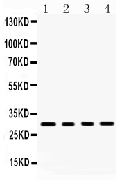
Figure 1. Western blot analysis of BAFF using anti-BAFF antibody (PB9567). Electrophoresis was performed on a 5-20% SDS-PAGE gel at 70V (Stacking gel) / 90V (Resolving gel) for 2-3 hours. Lane 1: Rat Kidney Tissue Lysate at 50ug, Lane 2: Rat Spleen Tissue Lysate at 50ug, Lane 3: Mouse Spleen Tissue Lysate at 50ug, Lane 4: Human Placenta Tissue Lysate at 50ug. After electrophoresis, proteins were transferred to a nitrocellulose membrane at 150 mA for 50-90 minutes. Blocked the membrane with 5% non-fat milk/TBS for 1.5 hour at RT. The membrane was incubated with rabbit anti-BAFF antigen affinity purified polyclonal antibody (Catalog # PB9567) at 0.5 microg/mL overnight at 4°C, then washed with TBS-0.1%Tween 3 times with 5 minutes each and probed with a goat anti-rabbit IgG-HRP secondary antibody at a dilution of 1:5000 for 1.5 hour at RT. The signal is developed using an Enhanced Chemiluminescent detection (ECL) kit (Catalog # EK1002) with Tanon 5200 system. A specific band was detected for BAFF at approximately 31 kDa. The expected band size for BAFF is at 31 kDa.
Figure 1. Western blot analysis of BAFF using anti-BAFF antibody (PB9567). Electrophoresis was performed on a 5-20% SDS-PAGE gel at 70V (Stacking gel) / 90V (Resolving gel) for 2-3 hours. Lane 1: Rat Kidney Tissue Lysate at 50ug, Lane 2: Rat Spleen Tissue Lysate at 50ug, Lane 3: Mouse Spleen Tissue Lysate at 50ug, Lane 4: Human Placenta Tissue Lysate at 50ug. After electrophoresis, proteins were transferred to a nitrocellulose membrane at 150 mA for 50-90 minutes. Blocked the membrane with 5% non-fat milk/TBS for 1.5 hour at RT. The membrane was incubated with rabbit anti-BAFF antigen affinity purified polyclonal antibody (Catalog # PB9567) at 0.5 microg/mL overnight at 4°C, then washed with TBS-0.1%Tween 3 times with 5 minutes each and probed with a goat anti-rabbit IgG-HRP secondary antibody at a dilution of 1:5000 for 1.5 hour at RT. The signal is developed using an Enhanced Chemiluminescent detection (ECL) kit (Catalog # EK1002) with Tanon 5200 system. A specific band was detected for BAFF at approximately 31 kDa. The expected band size for BAFF is at 31 kDa.
Anti-BAFF/TNFSF13B Antibody Picoband(r)

PB9567
ApplicationsWestern Blot
Product group Antibodies
ReactivityHuman, Mouse, Rat
TargetTNFSF13B
Overview
- SupplierBoster Bio
- Product NameAnti-BAFF Picoband Antibody
- Delivery Days Customer9
- Antibody SpecificityNo cross reactivity with other proteins.
- Application Supplier NoteTested Species: In-house tested species with positive results. Other applications have not been tested. Optimal dilutions should be determined by end users.
- ApplicationsWestern Blot
- CertificationResearch Use Only
- ClonalityPolyclonal
- Concentration500 ug/ml
- FormulationLyophilized
- Gene ID10673
- Target nameTNFSF13B
- Target descriptionTNF superfamily member 13b
- Target synonymsApoL related ligand TALL-1; BAFF; B-cell-activating factor; B-lymphocyte stimulator; BLYS; CD257; delta BAFF; Delta4 BAFF; dendritic cell-derived TNF-like molecule; DTL; epididymis secretory sperm binding protein; TALL1; TALL-1; THANK; TNF and ApoL-related leukocyte expressed ligand 1; TNF homolog that activates apoptosis; TNFSF20; TNLG7A; tumor necrosis factor (ligand) superfamily, member 13b; tumor necrosis factor (ligand) superfamily, member 20; tumor necrosis factor ligand 7A; tumor necrosis factor ligand superfamily member 13B; tumor necrosis factor superfamily member 13b; tumor necrosis factor-like protein ZTNF4; ZTNF4
- HostRabbit
- IsotypeIgG
- Protein IDQ9Y275
- Protein NameTumor necrosis factor ligand superfamily member 13B
- Scientific DescriptionBoster Bio Anti-BAFF/TNFSF13B Antibody Picoband® catalog # PB9567. Tested in WB applications. This antibody reacts with Human, Mouse, Rat. The brand Picoband indicates this is a premium antibody that guarantees superior quality, high affinity, and strong signals with minimal background in Western blot applications. Only our best-performing antibodies are designated as Picoband, ensuring unmatched performance.
- ReactivityHuman, Mouse, Rat
- Storage Instruction-20°C,2°C to 8°C
- UNSPSC12352203