

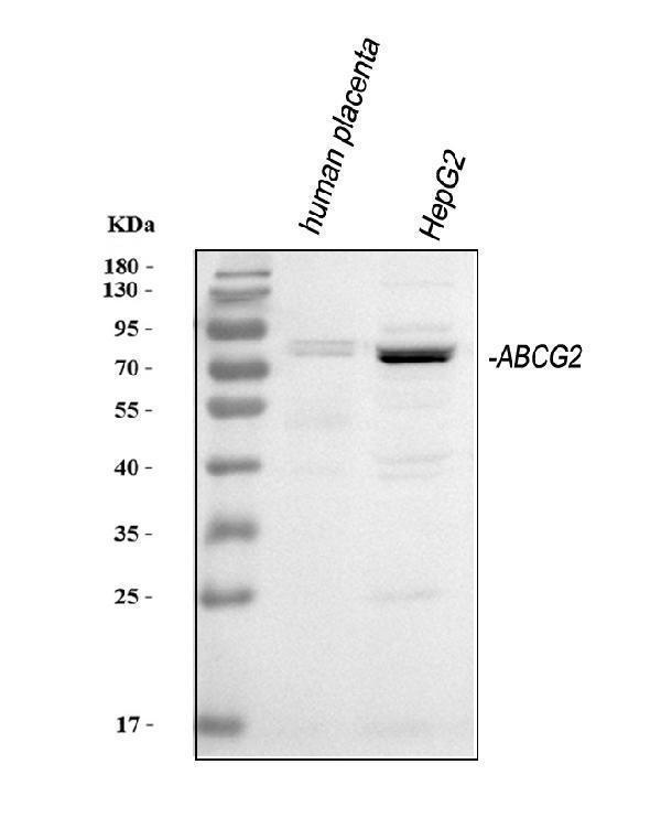
Figure 1. Western blot analysis of BCRP/ABCG2 using anti-BCRP/ABCG2 antibody (PA1869). Electrophoresis was performed on a 5-20% SDS-PAGE gel at 70V (Stacking gel) / 90V (Resolving gel) for 2-3 hours. The sample well of each lane was loaded with 30 ug of sample under reducing conditions. Lane 1: human placenta tissue lysates, Lane 2: human HepG2 whole cell lysates. After electrophoresis, proteins were transferred to a nitrocellulose membrane at 150 mA for 50-90 minutes. Blocked the membrane with 5% non-fat milk/TBS for 1.5 hour at RT. The membrane was incubated with rabbit anti-BCRP/ABCG2 antigen affinity purified polyclonal antibody (Catalog # PA1869) at 0.5 microg/mL overnight at 4°C, then washed with TBS-0.1%Tween 3 times with 5 minutes each and probed with a goat anti-rabbit IgG-HRP secondary antibody at a dilution of 1:5000 for 1.5 hour at RT. The signal is developed using an Enhanced Chemiluminescent detection (ECL) kit (Catalog # EK1002) with Tanon 5200 system. A specific band was detected for BCRP/ABCG2 at approximately 75 kDa. The expected band size for BCRP/ABCG2 is at 72 kDa.
Figure 1. Western blot analysis of BCRP/ABCG2 using anti-BCRP/ABCG2 antibody (PA1869). Electrophoresis was performed on a 5-20% SDS-PAGE gel at 70V (Stacking gel) / 90V (Resolving gel) for 2-3 hours. The sample well of each lane was loaded with 30 ug of sample under reducing conditions. Lane 1: human placenta tissue lysates, Lane 2: human HepG2 whole cell lysates. After electrophoresis, proteins were transferred to a nitrocellulose membrane at 150 mA for 50-90 minutes. Blocked the membrane with 5% non-fat milk/TBS for 1.5 hour at RT. The membrane was incubated with rabbit anti-BCRP/ABCG2 antigen affinity purified polyclonal antibody (Catalog # PA1869) at 0.5 microg/mL overnight at 4°C, then washed with TBS-0.1%Tween 3 times with 5 minutes each and probed with a goat anti-rabbit IgG-HRP secondary antibody at a dilution of 1:5000 for 1.5 hour at RT. The signal is developed using an Enhanced Chemiluminescent detection (ECL) kit (Catalog # EK1002) with Tanon 5200 system. A specific band was detected for BCRP/ABCG2 at approximately 75 kDa. The expected band size for BCRP/ABCG2 is at 72 kDa.
Anti-BCRP/ABCG2 Antibody Picoband(r)

PA1869
ApplicationsWestern Blot
Product group Antibodies
ReactivityHuman
TargetABCG2
Overview
- SupplierBoster Bio
- Product NameAnti-BCRP/ABCG2 Antibody
- Delivery Days Customer9
- Antibody SpecificityNo cross reactivity with other proteins.
- Application Supplier NoteWB: The detection limit for ABCG2 is approximately 1ng/lane under reducing conditions. Tested Species: In-house tested species with positive results. Predicted Species: Species predicted to be fit for the product based on sequence similarities. Other applications have not been tested. Optimal dilutions should be determined by end users.
- ApplicationsWestern Blot
- Applications SupplierWB
- CertificationResearch Use Only
- ClonalityPolyclonal
- Concentration500 ug/ml
- FormulationLyophilized
- Gene ID9429
- Target nameABCG2
- Target descriptionATP binding cassette subfamily G member 2 (Junior blood group)
- Target synonymsABC transporter; ABC15; ABCP; ATP-binding cassette transporter G2; ATP-binding cassette, sub-family G (WHITE), member 2 (Junior blood group); BCRP; BCRP1; BMDP; breast cancer resistance protein; broad substrate specificity ATP-binding cassette transporter ABCG2; CD338; CDw338; EST157481; GOUT1; mitoxantrone resistance-associated protein; MRX; multi drug resistance efflux transport ATP-binding cassette sub-family G (WHITE) member 2; MXR; MXR1; MXR-1; placenta specific MDR protein; placenta-specific ATP-binding cassette transporter; UAQTL1; urate exporter
- HostRabbit
- IsotypeIgG
- Protein IDQ9UNQ0
- Protein NameATP-binding cassette sub-family G member 2
- Scientific DescriptionBoster Bio Anti-BCRP/ABCG2 Antibody catalog # PA1869. Tested in WB applications. This antibody reacts with Human. The brand Picoband indicates this is a premium antibody that guarantees superior quality, high affinity, and strong signals with minimal background in Western blot applications. Only our best-performing antibodies are designated as Picoband, ensuring unmatched performance.
- ReactivityHuman
- Reactivity SupplierHuman
- Storage Instruction-20°C,2°C to 8°C
- UNSPSC12352203