

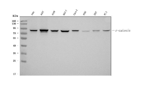
Figure 1. Western blot analysis of Catenin gamma(Plakoglobin) using anti-Catenin gamma(Plakoglobin) antibody (MA1012). Electrophoresis was performed on a 5-20% SDS-PAGE gel at 70V (Stacking gel) / 90V (Resolving gel) for 2-3 hours. The sample well of each lane was loaded with 30 ug of sample under reducing conditions. Lane 1: human Hela whole cell lysates, Lane 2: human A431 whole cell lysates, Lane 3: human A549 whole cell lysates, Lane 4: human MCF-7 whole cell lysates, Lane 5: human CACO-2 whole cell lysates, Lane 6: human K562 whole cell lysates, Lane 7: human 293T whole cell lysates, Lane 8: human PC-3 whole cell lysates. After electrophoresis, proteins were transferred to a nitrocellulose membrane at 150 mA for 50-90 minutes. Blocked the membrane with 5% non-fat milk/TBS for 1.5 hour at RT. The membrane was incubated with mouse anti-Catenin gamma(Plakoglobin) antigen affinity purified monoclonal antibody (Catalog # MA1012) at 1 microg/mL overnight at 4°C, then washed with TBS-0.1%Tween 3 times with 5 minutes each and probed with a goat anti-mouse IgG-HRP secondary antibody at a dilution of 1:10000 for 1.5 hour at RT. The signal is developed using an Enhanced Chemiluminescent detection (ECL) kit (Catalog # EK1001) with Tanon 5200 system. A specific band was detected for Catenin gamma(Plakoglobin) at approximately 82 kDa. The expected band size for Catenin gamma(Plakoglobin) is at 82 kDa.
Figure 1. Western blot analysis of Catenin gamma(Plakoglobin) using anti-Catenin gamma(Plakoglobin) antibody (MA1012). Electrophoresis was performed on a 5-20% SDS-PAGE gel at 70V (Stacking gel) / 90V (Resolving gel) for 2-3 hours. The sample well of each lane was loaded with 30 ug of sample under reducing conditions. Lane 1: human Hela whole cell lysates, Lane 2: human A431 whole cell lysates, Lane 3: human A549 whole cell lysates, Lane 4: human MCF-7 whole cell lysates, Lane 5: human CACO-2 whole cell lysates, Lane 6: human K562 whole cell lysates, Lane 7: human 293T whole cell lysates, Lane 8: human PC-3 whole cell lysates. After electrophoresis, proteins were transferred to a nitrocellulose membrane at 150 mA for 50-90 minutes. Blocked the membrane with 5% non-fat milk/TBS for 1.5 hour at RT. The membrane was incubated with mouse anti-Catenin gamma(Plakoglobin) antigen affinity purified monoclonal antibody (Catalog # MA1012) at 1 microg/mL overnight at 4°C, then washed with TBS-0.1%Tween 3 times with 5 minutes each and probed with a goat anti-mouse IgG-HRP secondary antibody at a dilution of 1:10000 for 1.5 hour at RT. The signal is developed using an Enhanced Chemiluminescent detection (ECL) kit (Catalog # EK1001) with Tanon 5200 system. A specific band was detected for Catenin gamma(Plakoglobin) at approximately 82 kDa. The expected band size for Catenin gamma(Plakoglobin) is at 82 kDa.
Anti-Catenin Gamma (Plakoglobin) Jup Antibody (Monoclonal, 15F11)
MA1012
ApplicationsWestern Blot, ImmunoCytoChemistry, ImmunoHistoChemistry
Product group Antibodies
ReactivityBovine, Human, Monkey
TargetJUP
Overview
- SupplierBoster Bio
- Product NameAnti-Catenin Gamma (Plakoglobin) Antibody (Monoclonal, 15F11)
- Delivery Days Customer9
- Antibody SpecificityNo cross reactivity with other proteins.
- Application Supplier NoteOther applications have not been tested. Optimal dilutions should be determined by end users.
- ApplicationsWestern Blot, ImmunoCytoChemistry, ImmunoHistoChemistry
- Applications SupplierIHF, ICC, WB, IHC
- CertificationResearch Use Only
- ClonalityMonoclonal
- Clone ID15F11
- Concentration100 ug/ml
- Gene ID3728
- Target nameJUP
- Target descriptionjunction plakoglobin
- Target synonymscatenin (cadherin-associated protein), gamma 80kDa; CTNNG; desmoplakin III; desmoplakin-3; desmosomal protein 3; DP3; DPIII; junction plakoglobin; PDGB; PG; PKGB
- HostMouse
- IsotypeIgG1
- Protein IDQ6P0K8
- Protein NameJunction plakoglobin
- Scientific DescriptionBoster Bio Anti-Catenin Gamma (Plakoglobin) Jup Antibody (Monoclonal, 15F11) catalog # MA1012. Tested in IHC, ICC, WB applications. This antibody reacts with Bovine, Human.
- ReactivityBovine, Human, Monkey
- Reactivity SupplierBovine, Human, Monkey
- Storage Instruction-20°C,2°C to 8°C
- UNSPSC12352203