

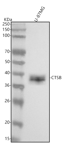
Figure 1. Western blot analysis of Cathepsin B using anti-Cathepsin B antibody (M01456). Electrophoresis was performed on a 5-20% SDS-PAGE gel at 70V (Stacking gel) / 90V (Resolving gel) for 2-3 hours. The sample well of each lane was loaded with 30 ug of sample under reducing conditions. Lane 1: human U-87MG whole cell lysates. After electrophoresis, proteins were transferred to a nitrocellulose membrane at 150 mA for 50-90 minutes. Blocked the membrane with 5% non-fat milk/TBS for 1.5 hour at RT. The membrane was incubated with rabbit anti-Cathepsin B antigen affinity purified monoclonal antibody (Catalog # M01456) at 1:500 overnight at 4°C, then washed with TBS-0.1%Tween 3 times with 5 minutes each and probed with a goat anti-rabbit IgG-HRP secondary antibody at a dilution of 1:1000 for 1.5 hour at RT. The signal is developed using an Enhanced Chemiluminescent detection (ECL) kit (Catalog # EK1002) with Tanon 5200 system. A specific band was detected for Cathepsin B at approximately 38 kDa. The expected band size for Cathepsin B is at 38 kDa.
. Cathepsin B was detected in a paraffin-embedded section of human colorectal adenocarcinoma tissue. Heat mediated antigen retrieval was performed in EDTA buffer (pH 8.0, epitope retrieval solution). The tissue section was blocked with 10% goat serum. The tissue section was then incubated with 1:50 rabbit anti-Cathepsin B Antibody M01456) overnight at 4°C. Peroxidase Conjugated Goat Anti-rabbit IgG was used as secondary antibody and incubated for 30 minutes at 37°C. The tissue section was developed using HRP Conjugated Rabbit IgG Super Vision Assay Kit (Catalog # SV0002) with DAB as the chromogen.")
. Cathepsin B was detected in a paraffin-embedded section of human liver cancer tissue. Heat mediated antigen retrieval was performed in EDTA buffer (pH 8.0, epitope retrieval solution). The tissue section was blocked with 10% goat serum. The tissue section was then incubated with 1:50 rabbit anti-Cathepsin B Antibody M01456) overnight at 4°C. Peroxidase Conjugated Goat Anti-rabbit IgG was used as secondary antibody and incubated for 30 minutes at 37°C. The tissue section was developed using HRP Conjugated Rabbit IgG Super Vision Assay Kit (Catalog # SV0002) with DAB as the chromogen.")
. Cathepsin B was detected in a paraffin-embedded section of human spleen tissue. Heat mediated antigen retrieval was performed in EDTA buffer (pH 8.0, epitope retrieval solution). The tissue section was blocked with 10% goat serum. The tissue section was then incubated with 1:50 rabbit anti-Cathepsin B Antibody M01456) overnight at 4°C. Peroxidase Conjugated Goat Anti-rabbit IgG was used as secondary antibody and incubated for 30 minutes at 37°C. The tissue section was developed using HRP Conjugated Rabbit IgG Super Vision Assay Kit (Catalog # SV0002) with DAB as the chromogen.")
Figure 1. Western blot analysis of Cathepsin B using anti-Cathepsin B antibody (M01456). Electrophoresis was performed on a 5-20% SDS-PAGE gel at 70V (Stacking gel) / 90V (Resolving gel) for 2-3 hours. The sample well of each lane was loaded with 30 ug of sample under reducing conditions. Lane 1: human U-87MG whole cell lysates. After electrophoresis, proteins were transferred to a nitrocellulose membrane at 150 mA for 50-90 minutes. Blocked the membrane with 5% non-fat milk/TBS for 1.5 hour at RT. The membrane was incubated with rabbit anti-Cathepsin B antigen affinity purified monoclonal antibody (Catalog # M01456) at 1:500 overnight at 4°C, then washed with TBS-0.1%Tween 3 times with 5 minutes each and probed with a goat anti-rabbit IgG-HRP secondary antibody at a dilution of 1:1000 for 1.5 hour at RT. The signal is developed using an Enhanced Chemiluminescent detection (ECL) kit (Catalog # EK1002) with Tanon 5200 system. A specific band was detected for Cathepsin B at approximately 38 kDa. The expected band size for Cathepsin B is at 38 kDa.
Anti-Cathepsin B CTSB Rabbit Monoclonal Antibody

M01456
ApplicationsImmunoFluorescence, ImmunoPrecipitation, Western Blot, ImmunoCytoChemistry, ImmunoHistoChemistry
Product group Antibodies
ReactivityHuman
TargetCTSB
Overview
- SupplierBoster Bio
- Product NameAnti-Cathepsin B Rabbit Monoclonal Antibody
- Delivery Days Customer9
- ApplicationsImmunoFluorescence, ImmunoPrecipitation, Western Blot, ImmunoCytoChemistry, ImmunoHistoChemistry
- CertificationResearch Use Only
- ClonalityMonoclonal
- Clone IDAAFO-3
- FormulationLiquid
- Gene ID1508
- Target nameCTSB
- Target descriptioncathepsin B
- Target synonymsamyloid precursor protein secretase; APP secretase; APPS; cathepsin B; cathepsin B1; CPSB; cysteine protease; epididymis secretory sperm binding protein; RECEUP
- HostRabbit
- IsotypeIgG
- Protein IDP07858
- Protein NameCathepsin B
- Scientific DescriptionBoster Bio Anti-Cathepsin B CTSB Rabbit Monoclonal Antibody catalog # M01456. Tested in WB, IHC, ICC/IF, IP applications. This antibody reacts with Human.
- ReactivityHuman
- Storage Instruction-20°C
- UNSPSC12352203
References
- The molecular mechanism for effects of TiN coating on NiTi alloy on endothelial cell function. Yang D et al., 2014 Aug, BiomaterialsRead more