


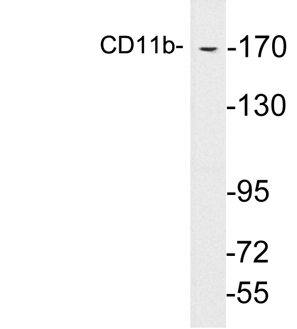
Figure 1. Western blot analysis of CD11b using anti-CD11b antibody (A00144-1). Electrophoresis was performed on a 5-20% SDS-PAGE gel at 70V (Stacking gel) / 90V (Resolving gel) for 2-3 hours. The sample well of each lane was loaded with 50ug of sample under reducing conditions. Lane 1: rat spleen tissue lysates, Lane 2: rat thymus tissue lysates, Lane 3: mouse spleen tissue lysates, Lane 4: mouse thymus tissue lysates. After Electrophoresis, proteins were transferred to a Nitrocellulose membrane at 150mA for 50-90 minutes. Blocked the membrane with 5% Non-fat Milk/ TBS for 1.5 hour at RT. The membrane was incubated with rabbit anti-CD11b antigen affinity purified polyclonal antibody (Catalog # A00144-1) at 0.5 microg/mL overnight at 4°C, then washed with TBS-0.1%Tween 3 times with 5 minutes each and probed with a goat anti-rabbit IgG-HRP secondary antibody at a dilution of 1:10000 for 1.5 hour at RT. The signal is developed using an Enhanced Chemiluminescent detection (ECL) kit (Catalog # EK1002) with Tanon 5200 system. A specific band was detected for CD11b at approximately 170KD. The expected band size for CD11b is at 127KD.
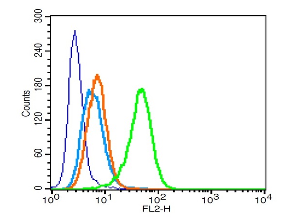
. Overlay histogram showing THP-1 cells stained with A00144-1 (Blue line). The cells were fixed with 4% paraformaldehyde and blocked with 10% normal goat serum. And then incubated with rabbit anti-CD11b Antibody (A00144-1, 1 microg/1x106 cells) for 30 min at 20°C. DyLight®488 conjugated goat anti-rabbit IgG (BA1127, 5-10 microg/1x106 cells) was used as secondary antibody for 30 minutes at 20°C. Isotype control antibody (Green line) was rabbit IgG (1 microg/1x106) used under the same conditions. Unlabelled sample without incubation with primary antibody and secondary antibody (Red line) was used as a blank control.")
Figure 1. Western blot analysis of CD11b using anti-CD11b antibody (A00144-1). Electrophoresis was performed on a 5-20% SDS-PAGE gel at 70V (Stacking gel) / 90V (Resolving gel) for 2-3 hours. The sample well of each lane was loaded with 50ug of sample under reducing conditions. Lane 1: rat spleen tissue lysates, Lane 2: rat thymus tissue lysates, Lane 3: mouse spleen tissue lysates, Lane 4: mouse thymus tissue lysates. After Electrophoresis, proteins were transferred to a Nitrocellulose membrane at 150mA for 50-90 minutes. Blocked the membrane with 5% Non-fat Milk/ TBS for 1.5 hour at RT. The membrane was incubated with rabbit anti-CD11b antigen affinity purified polyclonal antibody (Catalog # A00144-1) at 0.5 microg/mL overnight at 4°C, then washed with TBS-0.1%Tween 3 times with 5 minutes each and probed with a goat anti-rabbit IgG-HRP secondary antibody at a dilution of 1:10000 for 1.5 hour at RT. The signal is developed using an Enhanced Chemiluminescent detection (ECL) kit (Catalog # EK1002) with Tanon 5200 system. A specific band was detected for CD11b at approximately 170KD. The expected band size for CD11b is at 127KD.
Anti-CD11b/ITGAM Antibody Picoband(r)
A00144-1
ApplicationsFlow Cytometry, Western Blot, ELISA, ImmunoHistoChemistry
Product group Antibodies
ReactivityHuman, Mouse
TargetITGAM
Overview
- SupplierBoster Bio
- Product NameAnti-CD11b/ITGAM Antibody Picoband(r)
- Delivery Days Customer9
- Application Supplier NoteTested Species: In-house tested species with positive results. Other applications have not been tested. Optimal dilutions should be determined by end users.
- ApplicationsFlow Cytometry, Western Blot, ELISA, ImmunoHistoChemistry
- CertificationResearch Use Only
- ClonalityPolyclonal
- Concentration500 ug/ml
- Gene ID3684
- Target nameITGAM
- Target descriptionintegrin subunit alpha M
- Target synonymsCD11B, CR3A, MAC-1, MAC1A, MO1A, SLEB6, integrin alpha-M, CD11 antigen-like family member B, CR-3 alpha chain, antigen CD11b (p170), cell surface glycoprotein MAC-1 subunit alpha, complement component 3 receptor 3 subunit, integrin, alpha M (complement component 3 receptor 3 subunit), leukocyte adhesion receptor MO1, macrophage antigen alpha polypeptide, macrophage-1 antigen alpha subunit, neutrophil adherence receptor alpha-M subunit
- HostRabbit
- IsotypeIgG
- Protein IDP11215
- Protein NameIntegrin alpha-M
- Scientific DescriptionBoster Bio Anti-CD11b/ITGAM Antibody Picoband® catalog # A00144-1. Tested in ELISA, Flow Cytometry, IHC, WB applications. This antibody reacts with Human, Mouse. The brand Picoband indicates this is a premium antibody that guarantees superior quality, high affinity, and strong signals with minimal background in Western blot applications. Only our best-performing antibodies are designated as Picoband, ensuring unmatched performance.
- ReactivityHuman, Mouse
- Storage Instruction-20°C,2°C to 8°C
- UNSPSC12352203
References
- Luo F, Li R, Zheng H, et al. Differentiation of Bone Mesenchymal Stem Cells Into Vascular Endothelial Cell-Like Cells Using Functionalized Single-Walled Carbon Nanotubes. Front Bioeng Biotechnol. 2022,10:913080. doi: 10.3389/fbioe.2022.913080Read this paper
Datasheet
MSDS
Related products
Product group Antibodies
Anti-CD11b AntibodyA99157
ApplicationsWestern Blot, ELISA
ReactivityHuman, Mouse
- SizePrice
Product group Antibodies

Anti-Integrin alpha-M [107]Ab00473-1.1
ApplicationsFlow Cytometry, ELISA
ReactivityHuman
TargetITGAM
- SizePrice
Product group Antibodies
Anti-ITGAM Antibody144-01581
ApplicationsWestern Blot, ImmunoHistoChemistry
ReactivityHuman, Mouse, Rat
TargetITGAM
- SizePrice

Product group Antibodies
Anti-ITGAM AntibodyAMAB90911
ApplicationsWestern Blot, ImmunoHistoChemistry
ReactivityHuman
TargetITGAM
- SizePrice
Product group Antibodies

ITGAM / CD11b Antibody (clone M1/70)LS-C763543
ApplicationsFlow Cytometry, ImmunoPrecipitation, ImmunoHistoChemistry
ReactivityMouse
TargetITGAM
- SizePrice
Product group Antibodies
Anti-CD11b/ITGAM Antibody Picoband(r)A00144-1-CARRIER-FREE
ApplicationsFlow Cytometry, Western Blot, ELISA, ImmunoHistoChemistry
ReactivityHuman, Mouse
TargetITGAM
- SizePrice
Product group Antibodies
References
CD11b Polyclonal AntibodyBS-1014R
ApplicationsFlow Cytometry, ImmunoFluorescence, Western Blot, ELISA, ImmunoCytoChemistry, ImmunoHistoChemistry, ImmunoHistoChemistry Frozen, ImmunoHistoChemistry Paraffin
ReactivityHuman, Mouse, Rat
TargetITGAM
- SizePrice
Product group Antibodies
ITGAM AntibodyCSB-PA011876LA01HU
ApplicationsImmunoFluorescence, ELISA, ImmunoHistoChemistry
ReactivityHuman
TargetITGAM
- SizePrice
Product group Antibodies

Goat anti-ITGAM / CD11BEB07825
ApplicationsImmunoFluorescence, ELISA, ImmunoHistoChemistry
ReactivityHuman
TargetITGAM
- SizePrice