


Figure 1. Western blot analysis of CD14 using anti-CD14 antibody (A00137). Electrophoresis was performed on a 5-20% SDS-PAGE gel at 70V (Stacking gel) / 90V (Resolving gel) for 2-3 hours. The sample well of each lane was loaded with 50ug of sample under reducing conditions. Lane 1: mouse thymus tissue lysates, Lane 2: mouse spleen tissue lysates. After Electrophoresis, proteins were transferred to a Nitrocellulose membrane at 150mA for 50-90 minutes. Blocked the membrane with 5% Non-fat Milk/ TBS for 1.5 hour at RT. The membrane was incubated with rabbit anti-CD14 antigen affinity purified polyclonal antibody (Catalog # A00137) at 0.5 ug/mL overnight at 4 then washed with TBS-0.1%Tween 3 times with 5 minutes each and probed with a goat anti-rabbit IgG-HRP secondary antibody at a dilution of 1:10000 for 1.5 hour at RT. The signal is developed using an Enhanced Chemiluminescent detection (ECL) kit (Catalog # EK1002) with Tanon 5200 system. A specific band was detected for CD14 at approximately 50KD. The expected band size for CD14 is at 40KD.
. CD14 was detected in paraffin-embedded section of mouse spleen tissue. Heat mediated antigen retrieval was performed in citrate buffer (pH6, epitope retrieval solution) for 20 mins. The tissue section was blocked with 10% goat serum. The tissue section was then incubated with 1ug/ml rabbit anti-CD14 Antibody (A00137) overnight at 4 Biotinylated goat anti-rabbit IgG was used as secondary antibody and incubated for 30 minutes at 37 The tissue section was developed using Strepavidin-Biotin-Complex (SABC)(Catalog # SA1022) with DAB as the chromogen.")
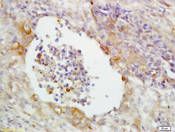
. CD14 was detected in paraffin-embedded section of rat lymphaden tissue. Heat mediated antigen retrieval was performed in citrate buffer (pH6, epitope retrieval solution) for 20 mins. The tissue section was blocked with 10% goat serum. The tissue section was then incubated with 1ug/ml rabbit anti-CD14 Antibody (A00137) overnight at 4 Biotinylated goat anti-rabbit IgG was used as secondary antibody and incubated for 30 minutes at 37 The tissue section was developed using Strepavidin-Biotin-Complex (SABC)(Catalog # SA1022) with DAB as the chromogen.")
. CD14 was detected in paraffin-embedded section of rat spleen tissue. Heat mediated antigen retrieval was performed in citrate buffer (pH6, epitope retrieval solution) for 20 mins. The tissue section was blocked with 10% goat serum. The tissue section was then incubated with 1ug/ml rabbit anti-CD14 Antibody (A00137) overnight at 4 Biotinylated goat anti-rabbit IgG was used as secondary antibody and incubated for 30 minutes at 37 The tissue section was developed using Strepavidin-Biotin-Complex (SABC)(Catalog # SA1022) with DAB as the chromogen.")
. Overlay histogram showing mouse PBMC cells stained with A00137 (Blue line). To facilitate intracellular staining, cells were fixed with 4% paraformaldehyde and permeabilized with permeabilization buffer. The cells were blocked with 10% normal goat serum. And then incubated with rabbit anti-CD14 Antibody (A00137,1microg/1x106 cells) for 30 min at 20°C. DyLight®488 conjugated goat anti-rabbit IgG (BA1127, 5-10microg/1x106 cells) was used as secondary antibody for 30 minutes at 20°C. Isotype control antibody (Green line) was rabbit IgG (1microg/1x106) used under the same conditions. Unlabelled sample without incubation with primary antibody and secondary antibody (Red line) was used as a blank control.")
Figure 1. Western blot analysis of CD14 using anti-CD14 antibody (A00137). Electrophoresis was performed on a 5-20% SDS-PAGE gel at 70V (Stacking gel) / 90V (Resolving gel) for 2-3 hours. The sample well of each lane was loaded with 50ug of sample under reducing conditions. Lane 1: mouse thymus tissue lysates, Lane 2: mouse spleen tissue lysates. After Electrophoresis, proteins were transferred to a Nitrocellulose membrane at 150mA for 50-90 minutes. Blocked the membrane with 5% Non-fat Milk/ TBS for 1.5 hour at RT. The membrane was incubated with rabbit anti-CD14 antigen affinity purified polyclonal antibody (Catalog # A00137) at 0.5 ug/mL overnight at 4 then washed with TBS-0.1%Tween 3 times with 5 minutes each and probed with a goat anti-rabbit IgG-HRP secondary antibody at a dilution of 1:10000 for 1.5 hour at RT. The signal is developed using an Enhanced Chemiluminescent detection (ECL) kit (Catalog # EK1002) with Tanon 5200 system. A specific band was detected for CD14 at approximately 50KD. The expected band size for CD14 is at 40KD.
Anti-CD14 Antibody
A00137
ApplicationsFlow Cytometry, Western Blot, ImmunoHistoChemistry
Product group Antibodies
ReactivityMouse, Rat
TargetCd14
Overview
- SupplierBoster Bio
- Product NameAnti-CD14 Antibody
- Delivery Days Customer9
- Application Supplier NoteTested Species: In-house tested species with positive results. By Heat: Boiling the paraffin sections in 10mM citrate buffer, pH6.0, for 20mins is required for the staining of formalin/paraffin sections. Other applications have not been tested. Optimal dilutions should be determined by end users.
- ApplicationsFlow Cytometry, Western Blot, ImmunoHistoChemistry
- CertificationResearch Use Only
- ClonalityPolyclonal
- Concentration500 ug/ml
- Gene ID12475
- Target nameCd14
- Target descriptionCD14 antigen
- Target synonymsmonocyte differentiation antigen CD14, myeloid cell-specific leucine-rich glycoprotein
- HostRabbit
- IsotypeIgG
- Protein IDP10810
- Protein NameMonocyte differentiation antigen CD14
- Scientific DescriptionBoster Bio Anti-CD14 Antibody Picoband® catalog # A00137. Tested in Flow Cytometry, IHC, WB applications. This antibody reacts with Mouse, Rat. The brand Picoband indicates this is a premium antibody that guarantees superior quality, high affinity, and strong signals with minimal background in Western blot applications. Only our best-performing antibodies are designated as Picoband, ensuring unmatched performance.
- ReactivityMouse, Rat
- Storage Instruction-20°C,2°C to 8°C
- UNSPSC12352203
Datasheet
MSDS
Related products
Product group Antibodies
ApplicationsFlow Cytometry
ReactivityMouse
TargetCd14
- SizePrice

Product group Antibodies
Anti-Cd14 Antibody Picoband(r)A00137-3-CARRIER-FREE
ApplicationsWestern Blot, ELISA
ReactivityMouse
TargetCd14
- SizePrice
Product group Antibodies
ApplicationsImmunoPrecipitation, Western Blot, ImmunoCytoChemistry, ImmunoHistoChemistry
ReactivityMouse
TargetCd14
- SizePrice
Product group Antibodies
References
CD14 Polyclonal AntibodyBS-1192R
ApplicationsImmunoFluorescence, Western Blot, ELISA, ImmunoCytoChemistry, ImmunoHistoChemistry, ImmunoHistoChemistry Frozen, ImmunoHistoChemistry Paraffin
ReactivityHuman, Mouse
TargetCd14
- SizePrice
![FACS analysis of 10? RAW264.7 cells using GTX54397 CD14 antibody [Sa14-2]. Black : No ab staining Red : Isotype control Blue : Primary antibody Dilution : 1μg/ml](https://www.genetex.com/upload/website/prouct_img/normal/GTX54397/GTX54397_20201207_FACS_5_w_23060900_581.webp)
Product group Antibodies
CD14 antibody [Sa14-2]GTX54397
ApplicationsFunctional Assay, Flow Cytometry, ImmunoFluorescence, ImmunoPrecipitation, Western Blot, ImmunoCytoChemistry
ReactivityMouse
TargetCd14
- SizePrice