

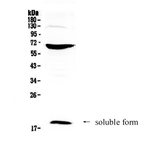
Figure 1. Western blot analysis of CD40L using anti-CD40L antibody (A01114-1). Electrophoresis was performed on a 5-20% SDS-PAGE gel at 70V (Stacking gel) / 90V (Resolving gel) for 2-3 hours. The sample well of each lane was loaded with 50ug of sample under reducing conditions. Lane 1: human HepG2 cell lysates. After Electrophoresis, proteins were transferred to a Nitrocellulose membrane at 150mA for 50-90 minutes. Blocked the membrane with 5% Non-fat Milk/ TBS for 1.5 hour at RT. The membrane was incubated with rabbit anti-CD40L antigen affinity purified polyclonal antibody (Catalog # A01114-1) at 0.5 microg/mL overnight at 4°C, then washed with TBS-0.1%Tween 3 times with 5 minutes each and probed with a goat anti-rabbit IgG-HRP secondary antibody at a dilution of 1:10000 for 1.5 hour at RT. The signal is developed using an Enhanced Chemiluminescent detection (ECL) kit (Catalog # EK1002) with Tanon 5200 system. A specific band was detected for CD40L at approximately 19KD. The expected band size for CD40L is at 29KD.
Figure 1. Western blot analysis of CD40L using anti-CD40L antibody (A01114-1). Electrophoresis was performed on a 5-20% SDS-PAGE gel at 70V (Stacking gel) / 90V (Resolving gel) for 2-3 hours. The sample well of each lane was loaded with 50ug of sample under reducing conditions. Lane 1: human HepG2 cell lysates. After Electrophoresis, proteins were transferred to a Nitrocellulose membrane at 150mA for 50-90 minutes. Blocked the membrane with 5% Non-fat Milk/ TBS for 1.5 hour at RT. The membrane was incubated with rabbit anti-CD40L antigen affinity purified polyclonal antibody (Catalog # A01114-1) at 0.5 microg/mL overnight at 4°C, then washed with TBS-0.1%Tween 3 times with 5 minutes each and probed with a goat anti-rabbit IgG-HRP secondary antibody at a dilution of 1:10000 for 1.5 hour at RT. The signal is developed using an Enhanced Chemiluminescent detection (ECL) kit (Catalog # EK1002) with Tanon 5200 system. A specific band was detected for CD40L at approximately 19KD. The expected band size for CD40L is at 29KD.
Anti-CD40L/CD40LG Antibody Picoband(r)
A01114-1-PE
ApplicationsWestern Blot, ELISA
Product group Antibodies
ReactivityHuman
TargetCD40LG
Overview
- SupplierBoster Bio
- Product NameAnti-CD40L/CD40LG Antibody Picoband(r)
- Delivery Days Customer9
- Antibody SpecificityNo cross reactivity with other proteins.
- ApplicationsWestern Blot, ELISA
- CertificationResearch Use Only
- ClonalityPolyclonal
- Concentration500 ug/ml
- ConjugateRPE
- Gene ID959
- Target nameCD40LG
- Target descriptionCD40 ligand
- Target synonymsCD154; CD40 antigen ligand; CD40 ligand; CD40L; CD40-L; gp39; hCD40L; HIGM1; IGM; IMD3; T-B cell-activating molecule; T-BAM; T-cell antigen Gp39; TNF-related activation protein; TNFSF5; TRAP; tumor necrosis factor (ligand) superfamily member 5
- HostRabbit
- IsotypeIgG
- Protein IDP29965
- Protein NameCD40 ligand
- Scientific DescriptionBoster Bio Anti-CD40L/CD40LG Antibody Picoband® catalog # A01114-1. Tested in ELISA, WB applications. This antibody reacts with Human. The brand Picoband indicates this is a premium antibody that guarantees superior quality, high affinity, and strong signals with minimal background in Western blot applications. Only our best-performing antibodies are designated as Picoband, ensuring unmatched performance.
- ReactivityHuman
- Storage Instruction-20°C,2°C to 8°C
- UNSPSC12352203