

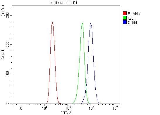
Figure 5. Flow Cytometry analysis of HL-60 cells using anti-CD44 antibody (PA1021-2). Overlay histogram showing HL-60 cells stained with PA1021-2 (Blue line). To facilitate intracellular staining, cells were fixed with 4% paraformaldehyde and permeabilized with permeabilization buffer. The cells were blocked with 10% normal goat serum. And then incubated with rabbit anti-CD44 Antibody (PA1021-2, 1 microg/1x106 cells) for 30 min at 20°C. DyLight®488 conjugated goat anti-rabbit IgG (BA1127, 5-10 microg/1x106 cells) was used as secondary antibody for 30 minutes at 20°C. Isotype control antibody (Green line) was rabbit IgG (1 microg/1x106) used under the same conditions. Unlabelled sample without incubation with primary antibody and secondary antibody (Red line) was used as a blank control.
. Electrophoresis was performed on a 5-20% SDS-PAGE gel at 70V (Stacking gel) / 90V (Resolving gel) for 2-3 hours. The sample well of each lane was loaded with 30 ug of sample under reducing conditions. Lane 1: human Hela whole cell lysates, Lane 2: rat PC-12 whole cell lysates. After electrophoresis, proteins were transferred to a nitrocellulose membrane at 150 mA for 50-90 minutes. Blocked the membrane with 5% non-fat milk/TBS for 1.5 hour at RT. The membrane was incubated with rabbit anti-CD44 antigen affinity purified polyclonal antibody (Catalog # PA1021-2) at 0.5 microg/mL overnight at 4°C, then washed with TBS-0.1%Tween 3 times with 5 minutes each and probed with a goat anti-rabbit IgG-HRP secondary antibody at a dilution of 1:5000 for 1.5 hour at RT. The signal is developed using an Enhanced Chemiluminescent detection (ECL) kit (Catalog # EK1002) with Tanon 5200 system. A specific band was detected for CD44 at approximately 82 kDa. The expected band size for CD44 is at 82 kDa.")
. Electrophoresis was performed on a 5-20% SDS-PAGE gel at 70V (Stacking gel) / 90V (Resolving gel) for 2-3 hours. The sample well of each lane was loaded with 30 ug of sample under reducing conditions. After electrophoresis, proteins were transferred to a nitrocellulose membrane at 150 mA for 50-90 minutes. Blocked the membrane with 5% milk in PBS/0.05% Tween-20 (5% milk/PBS/Tw) for 1.5 hour at RT. The membrane was incubated with rabbit anti-CD44 antigen CD44 antibody (PA1021-2) at 1 ug/ml in 5% milk/BPS/Tw overnight at 4°C, then washed with TBS-0.1%Tween 3 times with 5 minutes each and probed with a goat anti-rabbit IgG-HRP secondary antibody at at 1:3,000 in 5% milk/PBS/Tw for 1.5 hour at RT. The signal is developed using a chemiluminescence: West Pico from Thermo Scientific. A specific band was detected for CD44 at approximately 81 kDa. The expected band size for CD44 is at 81 kDa.")
. CD44 was detected in a paraffin-embedded section of human laryngeal squamous cell carcinoma tissue. Heat mediated antigen retrieval was performed in EDTA buffer (pH 8.0, epitope retrieval solution). The tissue section was blocked with 10% goat serum. The tissue section was then incubated with 2 microg/ml rabbit anti-CD44 Antibody (PA1021-2) overnight at 4°C. Peroxidase Conjugated Goat Anti-rabbit IgG was used as secondary antibody and incubated for 30 minutes at 37°C. The tissue section was developed using HRP Conjugated Rabbit IgG Super Vision Assay Kit (Catalog # SV0002) with DAB as the chromogen.")
. CD44 was detected in a paraffin-embedded section of human rectal cancer tissue. Heat mediated antigen retrieval was performed in EDTA buffer (pH 8.0, epitope retrieval solution). The tissue section was blocked with 10% goat serum. The tissue section was then incubated with 2 microg/ml rabbit anti-CD44 Antibody (PA1021-2) overnight at 4°C. Peroxidase Conjugated Goat Anti-rabbit IgG was used as secondary antibody and incubated for 30 minutes at 37°C. The tissue section was developed using HRP Conjugated Rabbit IgG Super Vision Assay Kit (Catalog # SV0002) with DAB as the chromogen.")
. CD44 was detected in a paraffin-embedded section of rat lung tissue. Heat mediated antigen retrieval was performed in EDTA buffer (pH 8.0, epitope retrieval solution). The tissue section was blocked with 10% goat serum. The tissue section was then incubated with 2 microg/ml rabbit anti-CD44 Antibody (PA1021-2) overnight at 4°C. Peroxidase Conjugated Goat Anti-rabbit IgG was used as secondary antibody and incubated for 30 minutes at 37°C. The tissue section was developed using HRP Conjugated Rabbit IgG Super Vision Assay Kit (Catalog # SV0002) with DAB as the chromogen.")
Figure 5. Flow Cytometry analysis of HL-60 cells using anti-CD44 antibody (PA1021-2). Overlay histogram showing HL-60 cells stained with PA1021-2 (Blue line). To facilitate intracellular staining, cells were fixed with 4% paraformaldehyde and permeabilized with permeabilization buffer. The cells were blocked with 10% normal goat serum. And then incubated with rabbit anti-CD44 Antibody (PA1021-2, 1 microg/1x106 cells) for 30 min at 20°C. DyLight®488 conjugated goat anti-rabbit IgG (BA1127, 5-10 microg/1x106 cells) was used as secondary antibody for 30 minutes at 20°C. Isotype control antibody (Green line) was rabbit IgG (1 microg/1x106) used under the same conditions. Unlabelled sample without incubation with primary antibody and secondary antibody (Red line) was used as a blank control.
Anti-CD44 antigen CD44 Antibody Picoband(r)

PA1021-2
ApplicationsFlow Cytometry, Western Blot, ImmunoHistoChemistry
Product group Antibodies
ReactivityHamster, Human, Rat
TargetCD44
Overview
- SupplierBoster Bio
- Product NameAnti-CD44 Antibody
- Delivery Days Customer9
- Antibody SpecificityNo cross reactivity with other proteins.
- ApplicationsFlow Cytometry, Western Blot, ImmunoHistoChemistry
- Applications SupplierWB, IHC-P, IHC-F, ICC
- CertificationResearch Use Only
- ClonalityPolyclonal
- Concentration500 ug/ml
- FormulationLyophilized
- Gene ID960
- Target nameCD44
- Target descriptionCD44 molecule (Indian blood group)
- Target synonymsCD44 antigen; CDW44; cell surface glycoprotein CD44; chondroitin sulfate proteoglycan 8; CSPG8; ECMR-III; epican; extracellular matrix receptor III; GP90 lymphocyte homing/adhesion receptor; HCELL; hematopoietic cell E- and L-selectin ligand; heparan sulfate proteoglycan; Hermes antigen; homing function and Indian blood group system; HUTCH-I; hyaluronate receptor; IN; Indian blood group antigen; LHR; MC56; MDU2; MDU3; MIC4; Pgp1; phagocytic glycoprotein 1; soluble CD44
- HostRabbit
- IsotypeIgG
- Protein IDP16070
- Protein NameCD44 antigen
- Scientific DescriptionBoster Bio Anti-CD44 antigen CD44 Antibody catalog # PA1021-2. Tested in Flow Cytometry, IHC, WB applications. This antibody reacts with Human, Rat. The brand Picoband indicates this is a premium antibody that guarantees superior quality, high affinity, and strong signals with minimal background in Western blot applications. Only our best-performing antibodies are designated as Picoband, ensuring unmatched performance.
- ReactivityHamster, Human, Rat
- Reactivity SupplierReacts with: human, mouse, rat
- Storage Instruction-20°C,2°C to 8°C
- UNSPSC12352203
References
- Rhus coriaria L. (Sumac) Demonstrates Oncostatic Activity in the Therapeutic and Preventive Model of Breast Carcinoma. Kubatka P et al., 2020 Dec 26, Int J Mol SciRead more