


Figure 1. Western blot analysis of COL1A1 using anti-COL1A1 antibody (PA2140-1). Electrophoresis was performed on a 5-20% SDS-PAGE gel at 70V (Stacking gel) / 90V (Resolving gel) for 2-3 hours. The sample well of each lane was loaded with 30 ug of sample under reducing conditions. Lane 1: human HK2 whole cell lysates, Lane 2: human HCC tissue lysates, Lane 3: mouse NIH/3T3 whole cell lysates. After electrophoresis, proteins were transferred to a nitrocellulose membrane at 150 mA for 50-90 minutes. Blocked the membrane with 5% non-fat milk/TBS for 1.5 hour at RT. The membrane was incubated with rabbit anti-COL1A1 antigen affinity purified polyclonal antibody (Catalog # PA2140-1) at 0.5 microg/mL overnight at 4°C, then washed with TBS-0.1%Tween 3 times with 5 minutes each and probed with a goat anti-rabbit IgG-HRP secondary antibody at a dilution of 1:5000 for 1.5 hour at RT. The signal is developed using an Enhanced Chemiluminescent detection (ECL) kit (Catalog # EK1002) with Tanon 5200 system. A specific band was detected for COL1A1 at approximately 180-200 kDa. The expected band size for COL1A1 is at 138 kDa.
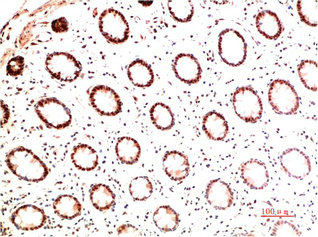
. COL1A1 was detected in a paraffin-embedded section of human colon adenocarcinoma tissue. Heat mediated antigen retrieval was performed in EDTA buffer (pH 8.0, epitope retrieval solution). The tissue section was blocked with 10% goat serum. The tissue section was then incubated with 2 microg/ml rabbit anti-COL1A1 Antibody (PA2140-1) overnight at 4°C. Biotinylated goat anti-rabbit IgG was used as secondary antibody and incubated for 30 minutes at 37°C. The tissue section was developed using Strepavidin-Biotin-Complex (SABC) (Catalog # SA1022) with DAB as the chromogen.")
. COL1A1 was detected in a paraffin-embedded section of human gastric carcinomaa tissue. Heat mediated antigen retrieval was performed in EDTA buffer (pH 8.0, epitope retrieval solution). The tissue section was blocked with 10% goat serum. The tissue section was then incubated with 2 microg/ml rabbit anti-COL1A1 Antibody (PA2140-1) overnight at 4°C. Biotinylated goat anti-rabbit IgG was used as secondary antibody and incubated for 30 minutes at 37°C. The tissue section was developed using Strepavidin-Biotin-Complex (SABC) (Catalog # SA1022) with DAB as the chromogen.")
. COL1A1 was detected in a paraffin-embedded section of human spleen tissue. Heat mediated antigen retrieval was performed in EDTA buffer (pH 8.0, epitope retrieval solution). The tissue section was blocked with 10% goat serum. The tissue section was then incubated with 2 microg/ml rabbit anti-COL1A1 Antibody (PA2140-1) overnight at 4°C. Biotinylated goat anti-rabbit IgG was used as secondary antibody and incubated for 30 minutes at 37°C. The tissue section was developed using Strepavidin-Biotin-Complex (SABC) (Catalog # SA1022) with DAB as the chromogen.")
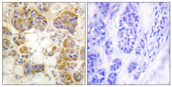
. COL1A1 was detected in a paraffin-embedded section of human endometrial cancer tissue. Heat mediated antigen retrieval was performed in EDTA buffer (pH 8.0, epitope retrieval solution). The tissue section was blocked with 10% goat serum. The tissue section was then incubated with 5 microg/mL rabbit anti-COL1A1 Antibody (PA2140-1) overnight at 4°C. DyLight®550 Conjugated Goat Anti-Rabbit IgG (BA1135) was used as secondary antibody at 1:500 dilution and incubated for 30 minutes at 37°C. The section was counterstained with DAPI. Visualize using a fluorescence microscope and filter sets appropriate for the label used.")
Figure 1. Western blot analysis of COL1A1 using anti-COL1A1 antibody (PA2140-1). Electrophoresis was performed on a 5-20% SDS-PAGE gel at 70V (Stacking gel) / 90V (Resolving gel) for 2-3 hours. The sample well of each lane was loaded with 30 ug of sample under reducing conditions. Lane 1: human HK2 whole cell lysates, Lane 2: human HCC tissue lysates, Lane 3: mouse NIH/3T3 whole cell lysates. After electrophoresis, proteins were transferred to a nitrocellulose membrane at 150 mA for 50-90 minutes. Blocked the membrane with 5% non-fat milk/TBS for 1.5 hour at RT. The membrane was incubated with rabbit anti-COL1A1 antigen affinity purified polyclonal antibody (Catalog # PA2140-1) at 0.5 microg/mL overnight at 4°C, then washed with TBS-0.1%Tween 3 times with 5 minutes each and probed with a goat anti-rabbit IgG-HRP secondary antibody at a dilution of 1:5000 for 1.5 hour at RT. The signal is developed using an Enhanced Chemiluminescent detection (ECL) kit (Catalog # EK1002) with Tanon 5200 system. A specific band was detected for COL1A1 at approximately 180-200 kDa. The expected band size for COL1A1 is at 138 kDa.
Anti-Collagen I Antibody
PA2140-1
ApplicationsWestern Blot, ImmunoHistoChemistry
Product group Antibodies
ReactivityHamster, Human, Mouse
TargetCOL1A1
Overview
- SupplierBoster Bio
- Product NameAnti-Collagen I Antibody
- Delivery Days Customer9
- ApplicationsWestern Blot, ImmunoHistoChemistry
- Applications SupplierIHP, IHF, ICC, WB, IHC
- CertificationResearch Use Only
- ClonalityPolyclonal
- Concentration500 ug/ml
- Gene ID1277
- Target nameCOL1A1
- Target descriptioncollagen type I alpha 1 chain
- Target synonymsCAFYD, EDSARTH1, EDSC, OI1, OI2, OI3, OI4, collagen alpha-1(I) chain, alpha-1 type I collagen, alpha1(I) procollagen, collagen Col1-ColIII-1, collagen Col1-ColIII-2, collagen alpha 1 chain type I, collagen alpha-1(I) chain preproprotein, collagen of skin, tendon and bone, alpha-1 chain, collagen, type I, alpha 1, pro-alpha-1 collagen type 1, type I proalpha 1, type I procollagen alpha 1 chain
- HostRabbit
- IsotypeIgG
- Protein IDP02452
- Protein NameCollagen alpha-1(I) chain
- Scientific DescriptionBoster Bio Anti-Collagen I/COL1A1 Antibody catalog # PA2140-1. Tested in IHC, WB applications. This antibody reacts with Human, Mouse The brand Picoband indicates this is a premium antibody that guarantees superior quality, high affinity, and strong signals with minimal background in Western blot applications. Only our best-performing antibodies are designated as Picoband, ensuring unmatched performance.
- ReactivityHamster, Human, Mouse
- Reactivity SupplierHuman, Mouse, Rat, Hamster
- Storage Instruction-20°C,2°C to 8°C
- UNSPSC12352203
References
- Zhang W, Peng Q, Huang X, et al. Commensal microbiome dysbiosis elicits interleukin-8 signaling to drive fibrotic skin disease. PNAS Nexus. 2024,3(7):pgae273. doi: 10.1093/pnasnexus/pgae273Read this paper
- Yan Y, Yan Q, Cai K, et al. Silk fibroin microgrooved zirconia surfaces improve connective tissue sealing through mediating glycolysis of fibroblasts. Mater Today Bio. 2024,27:101158. doi: 10.1016/j.mtbio.2024.101158Read this paper
- Li Y, Chen S, Yang Q, et al. The ANGPTL4-HIF-1α loop: a critical regulator of renal interstitial fibrosis. J Transl Med. 2024,22(1):649. doi: 10.1186/s12967-024-05466-3Read this paper
- Li G, Lu J, Wang C, et al. Discovery of Sophoridine α-Aryl Propionamide Derivative ZM600 as a Novel Antihepatic Fibrosis Agent. J Med Chem. 2024,67(13):11389-11400. doi: 10.1021/acs.jmedchem.4c01010Read this paper
- Chen ZY, Panga MJ, Zhang X, et al. Estrogen alleviates liver fibrosis and restores metabolic homeostasis in ovariectomy-induced liver injury and carbon tetrachloride (CCl(4)) exposure. Eur J Pharmacol. 2024,978:176774. doi: 10.1016/j.ejphar.2024.176774Read this paper
- Gong Y, Wang J, Pan M, et al. Harmine inhibits pulmonary fibrosis through regulating DNA damage repair-related genes and activation of TP53-Gadd45α pathway. Int Immunopharmacol. 2024,138:112542. doi: 10.1016/j.intimp.2024.112542Read this paper
- Gou Y, Li H, Sun X, et al. Parathyroid hormone (1-34) retards the lumbar facet joint degeneration and activates Wnt/β-catenin signaling pathway in ovariectomized rats. J Orthop Surg Res. 2024,19(1):352. doi: 10.1186/s13018-024-04817-6Read this paper
- Liang Z, Tang Z, Zhu C, et al. Intestinal CXCR6(+) ILC3s migrate to the kidney and exacerbate renal fibrosis via IL-23 receptor signaling enhanced by PD-1 expression. Immunity. 2024,57(6):1306-1323.e8. doi: 10.1016/j.immuni.2024.05.004Read this paper
- Xiong YB, Huang WY, Ling X, et al. Mitochondrial calcium uniporter promotes kidney aging in mice through inducing mitochondrial calcium-mediated renal tubular cell senescence. Acta Pharmacol Sin. 2024,45(10):2149-2162. doi: 10.1038/s41401-024-01298-5Read this paper
- Kang Z, Wang C, Shao F, et al. The increase of long noncoding RNA Fendrr in hepatocytes contributes to liver fibrosis by promoting IL-6 production. J Biol Chem. 2024,300(6):107376. doi: 10.1016/j.jbc.2024.107376Read this paper
Datasheet
MSDS
Related products
Product group Antibodies

Anti-collagen type I [3P1-31], Human IgG1-Fc Fusion,AB04222-10.159
ApplicationsELISA
ReactivityHuman
TargetCOL1A1
- SizePrice
Product group Antibodies
Anti-COL1A1 Antibody144-62810
ApplicationsImmunoFluorescence, ImmunoPrecipitation, Western Blot, ImmunoHistoChemistry
ReactivityHuman, Mouse, Rat
TargetCOL1A1
- SizePrice
Product group Antibodies

Collagen Type I, humanCO20111-0.1
ApplicationsImmunoFluorescence, Western Blot, ELISA, ImmunoHistoChemistry, ImmunoHistoChemistry Paraffin, RadioImmunoAssay
TargetCOL1A1
- SizePrice
Product group Antibodies
COL1A1 Monoclonal AntibodyCSB-MA139659
ApplicationsELISA, ImmunoHistoChemistry
ReactivityHuman, Mouse, Rat
TargetCOL1A1
- SizePrice
Product group Antibodies
References
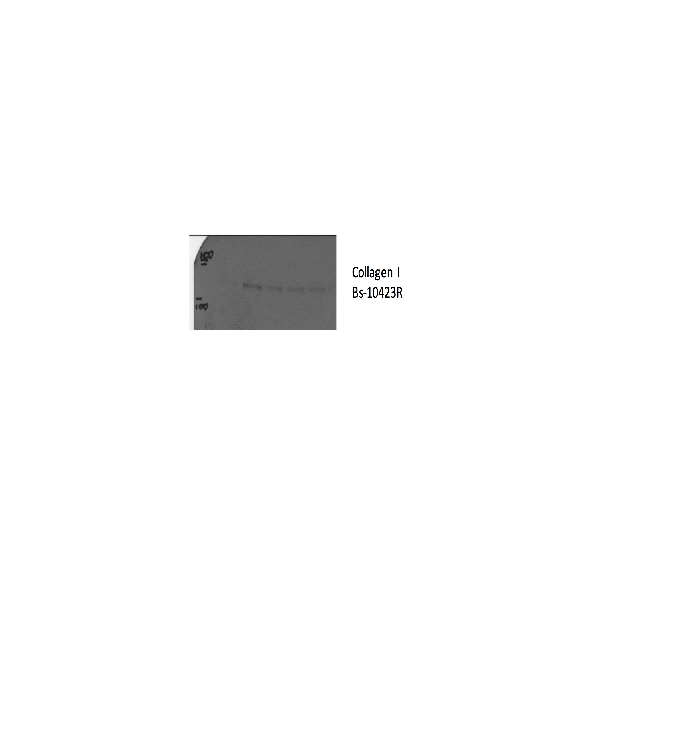
Collagen I Polyclonal AntibodyBS-10423R
ApplicationsImmunoFluorescence, Western Blot, ImmunoCytoChemistry, ImmunoHistoChemistry, ImmunoHistoChemistry Frozen, ImmunoHistoChemistry Paraffin
TargetCOL1A1
- SizePrice
Product group Antibodies
Anti-Collagen I AntibodyA95083
ApplicationsImmunoFluorescence, ELISA, ImmunoHistoChemistry
ReactivityHuman, Mouse, Rat
- SizePrice
Product group Antibodies

COL1A1 / Collagen I Alpha 1 AntibodyLS-C831483
ApplicationsImmunoHistoChemistry
ReactivityHuman
TargetCOL1A1
- SizePrice

Product group Antibodies
Anti-COL1A1 AntibodyHPA008405
ApplicationsImmunoHistoChemistry
ReactivityHuman
TargetCOL1A1
- SizePrice
Product group Antibodies
ApplicationsFlow Cytometry
TargetCOL1A1
- SizePrice
Product group Antibodies
COL1A2/COL1A1 AntibodyPACO07262
ApplicationsWestern Blot, ELISA
ReactivityHuman, Mouse, Rat
TargetCOL1A1
- SizePrice