

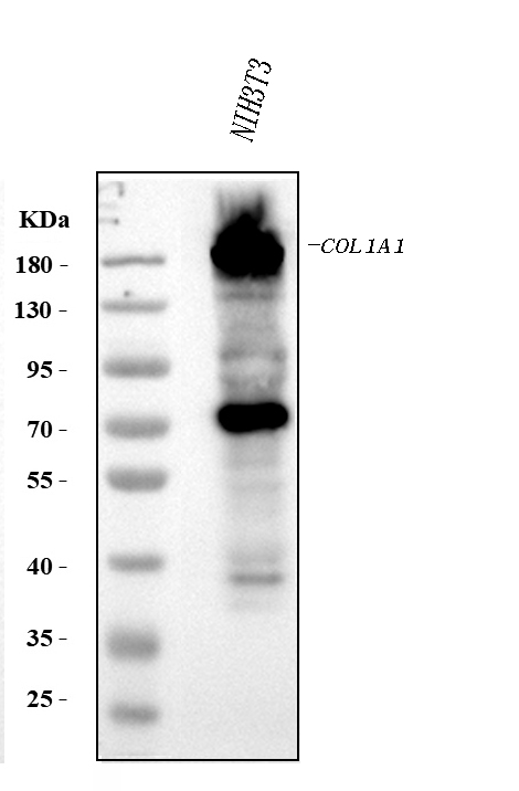
Figure 1. Western blot analysis of COL1A1 using anti-COL1A1 antibody (PB9939). Electrophoresis was performed on a 5-20% SDS-PAGE gel at 70V (Stacking gel) / 90V (Resolving gel) for 2-3 hours. The sample well of each lane was loaded with 30 ug of sample under reducing conditions. Lane 1: mouse NIH/3T3 whole cell lysates. After electrophoresis, proteins were transferred to a nitrocellulose membrane at 150 mA for 50-90 minutes. Blocked the membrane with 5% non-fat milk/TBS for 1.5 hour at RT. The membrane was incubated with rabbit anti-COL1A1 antigen affinity purified polyclonal antibody (Catalog # PB9939) at 0.5 microg/mL overnight at 4°C, then washed with TBS-0.1%Tween 3 times with 5 minutes each and probed with a goat anti-rabbit IgG-HRP secondary antibody at a dilution of 1:5000 for 1.5 hour at RT. The signal is developed using an Enhanced Chemiluminescent detection (ECL) kit (Catalog # EK1002) with Tanon 5200 system. A specific band was detected for COL1A1 at approximately 138-180 kDa. The expected band size for COL1A1 is at 138 kDa.
. COL1A1 was detected in a paraffin-embedded section of mouse lung tissue. Heat mediated antigen retrieval was performed in EDTA buffer (pH 8.0, epitope retrieval solution). The tissue section was blocked with 10% goat serum. The tissue section was then incubated with 2 microg/ml rabbit anti-COL1A1 Antibody (PB9939) overnight at 4°C. Peroxidase Conjugated Goat Anti-rabbit IgG was used as secondary antibody and incubated for 30 minutes at 37°C. The tissue section was developed using HRP Conjugated Rabbit IgG Super Vision Assay Kit (Catalog # SV0002) with DAB as the chromogen.")
. COL1A1 was detected in a paraffin-embedded section of rat lung tissue. Heat mediated antigen retrieval was performed in EDTA buffer (pH 8.0, epitope retrieval solution). The tissue section was blocked with 10% goat serum. The tissue section was then incubated with 2 microg/ml rabbit anti-COL1A1 Antibody (PB9939) overnight at 4°C. Peroxidase Conjugated Goat Anti-rabbit IgG was used as secondary antibody and incubated for 30 minutes at 37°C. The tissue section was developed using HRP Conjugated Rabbit IgG Super Vision Assay Kit (Catalog # SV0002) with DAB as the chromogen.")
Figure 1. Western blot analysis of COL1A1 using anti-COL1A1 antibody (PB9939). Electrophoresis was performed on a 5-20% SDS-PAGE gel at 70V (Stacking gel) / 90V (Resolving gel) for 2-3 hours. The sample well of each lane was loaded with 30 ug of sample under reducing conditions. Lane 1: mouse NIH/3T3 whole cell lysates. After electrophoresis, proteins were transferred to a nitrocellulose membrane at 150 mA for 50-90 minutes. Blocked the membrane with 5% non-fat milk/TBS for 1.5 hour at RT. The membrane was incubated with rabbit anti-COL1A1 antigen affinity purified polyclonal antibody (Catalog # PB9939) at 0.5 microg/mL overnight at 4°C, then washed with TBS-0.1%Tween 3 times with 5 minutes each and probed with a goat anti-rabbit IgG-HRP secondary antibody at a dilution of 1:5000 for 1.5 hour at RT. The signal is developed using an Enhanced Chemiluminescent detection (ECL) kit (Catalog # EK1002) with Tanon 5200 system. A specific band was detected for COL1A1 at approximately 138-180 kDa. The expected band size for COL1A1 is at 138 kDa.
Anti-Collagen I/COL1A1 Antibody Picoband(r)

PB9939
ApplicationsWestern Blot, ImmunoHistoChemistry
Product group Antibodies
ReactivityHamster, Mouse, Rat
TargetCol1a1
Overview
- SupplierBoster Bio
- Product NameAnti-Collagen I Picoband Antibody
- Delivery Days Customer9
- Antibody SpecificityNo cross reactivity with other proteins.
- Application Supplier NoteTested Species: In-house tested species with positive results. By Heat: Boiling the paraffin sections in 10mM citrate buffer, pH6.0, for 20mins is required for the staining of formalin/paraffin sections. Other applications have not been tested. Optimal dilutions should be determined by end users.
- ApplicationsWestern Blot, ImmunoHistoChemistry
- CertificationResearch Use Only
- ClonalityPolyclonal
- Concentration500 ug/ml
- FormulationLyophilized
- Gene ID12842
- Target nameCol1a1
- Target descriptioncollagen, type I, alpha 1
- Target synonymsalpha-1 type 1 collagen; alpha-1 type I collagen; Co; Col; Col1; Col1a-1; Cola1; Cola-1; collagen alpha-1(I) chain; Mov-; Mov13; Mov-13; procollagen, type I, alpha 1
- HostRabbit
- IsotypeIgG
- Protein IDP11087
- Protein NameCollagen alpha-1(I) chain
- Scientific DescriptionBoster Bio Anti-Collagen I/COL1A1 Antibody Picoband® catalog # PB9939. Tested in IHC, WB applications. This antibody reacts with Mouse, Rat. The brand Picoband indicates this is a premium antibody that guarantees superior quality, high affinity, and strong signals with minimal background in Western blot applications. Only our best-performing antibodies are designated as Picoband, ensuring unmatched performance.
- ReactivityHamster, Mouse, Rat
- Storage Instruction-20°C,2°C to 8°C
- UNSPSC12352203
References
- TH5487, a small molecule inhibitor of OGG1, attenuates pulmonary fibrosis by NEDD4L-mediated OGG1 degradation. Ling H et al., 2022 Aug 1, Chem Biol InteractRead more
- Visualized procollagen Ialpha1 demonstrates the intracellular processing of propeptides. Tanaka T et al., 2022 May, Life Sci AllianceRead more
- Microcystin-leucine arginine (MC-LR) induces mouse ovarian inflammation by promoting granulosa cells to produce inflammatory cytokine via activation of cGAS-STING signaling. Liu K et al., 2022 Apr 1, Toxicol LettRead more
- Genetic and pharmacological inhibition of fatty acid-binding protein 4 alleviated inflammation and early fibrosis after toxin induced kidney injury. Li L et al., 2021 Jul, Int ImmunopharmacolRead more
- Effect of Acellular Amnion With Increased TGF-beta and bFGF Levels on the Biological Behavior of Tenocytes. Sang R et al., 2020, Front Bioeng BiotechnolRead more
- Effective-component compatibility of Bufei Yishen formula II inhibits mucus hypersecretion of chronic obstructive pulmonary disease rats by regulating EGFR/PI3K/mTOR signaling. Li J et al., 2020 Jul 15, J EthnopharmacolRead more
- Tanshinone IIA attenuates silica-induced pulmonary fibrosis via inhibition of TGF-beta1-Smad signaling pathway. Feng F et al., 2020 Jan, Biomed PharmacotherRead more
- Exosomes Derived from Human Umbilical Cord Mesenchymal Stem Cells Promote Fibroblast-to-Myofibroblast Differentiation in Inflammatory Environments and Benefit Cardioprotective Effects. Shi Y et al., 2019 Jun 15, Stem Cells DevRead more
- Astragaloside exerts anti-photoaging effects in UVB-induced premature senescence of rat dermal fibroblasts through enhanced autophagy. Wen W et al., 2018 Nov 1, Arch Biochem BiophysRead more
- Co-culture with TM4 cells enhances the proliferation and migration of rat adipose-derived mesenchymal stem cells with high stemness. Luo Y et al., 2018 Oct, CytotechnologyRead more