

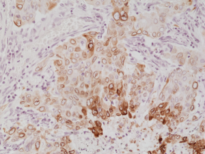
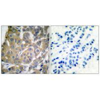
Immunohistochemical staining of formalin fixed and paraffin embedded human lung cancer tissue section using anti-COX-2 rabbit monoclonal antibody (Clone RM348) at a 1:1000 dilution.
Immunohistochemical staining of formalin fixed and paraffin embedded human lung cancer tissue section using anti-COX-2 rabbit monoclonal antibody (Clone RM348) at a 1:1000 dilution.
anti-COX-2 (human), Rabbit Monoclonal (RM348)
REV-31-1234-00
ApplicationsWestern Blot, ImmunoHistoChemistry
Product group Antibodies
ReactivityHuman
TargetPTGS2
Overview
- SupplierRevMAb Biosciences
- Product Nameanti-COX-2 (human), Rabbit Monoclonal (RM348)
- Delivery Days Customer10
- ApplicationsWestern Blot, ImmunoHistoChemistry
- CertificationResearch Use Only
- ClonalityMonoclonal
- Clone IDRM348
- Gene ID5743
- Target namePTGS2
- Target descriptionprostaglandin-endoperoxide synthase 2
- Target synonymsCOX-2, COX2, GRIPGHS, PGG/HS, PGHS-2, PHS-2, hCox-2, prostaglandin G/H synthase 2, PGH synthase 2, PHS II, cyclooxygenase 2, cyclooxygenase 2b, prostaglandin H2 synthase 2, prostaglandin-endoperoxide synthase 2 (prostaglandin G/H synthase and cyclooxygenase)
- HostRabbit
- IsotypeIgG
- Protein IDP35354
- Protein NameProstaglandin G/H synthase 2
- Scientific DescriptionCOX-2 converts arachidonate to prostaglandin H2 (PGH2), a committed step in prostanoid synthesis, including production of inflammatory prostaglandins. The conversion of arachidonate to prostaglandin H2 is a 2 step reaction: a cyclooxygenase (COX) reaction which converts arachidonate to prostaglandin G2 (PGG2) and a peroxidase reaction in which PGG2 is reduced to prostaglandin H2 (PGH2). COX-2 is constitutively expressed in some tissues in physiological conditions, such as the endothelium, kidney and brain, and is up-regulated under pathological conditions, such as in cancer and inflammation. Up-regulation of COX2 is also associated with increased cell adhesion, phenotypic changes, resistance to apoptosis and tumor angiogenesis. In cancer cells, COX-2 is a key step in the production of prostaglandin E2 (PGE2), which plays important roles in modulating motility, proliferation and resistance to apoptosis. COX-2 is naturally inhibited by calcitriol (the active form of Vitamin D). COX-2 is a target of NSAID such as aspirin, which can reduce pain and swelling from inflammation driven by COX-2. - Recombinant Antibody. This antibody reacts to human COX-2 (Prostaglandin G/H synthase 2). Applications: WB, IHC. Source: Rabbit. Liquid. 50% Glycerol/PBS with 1% BSA and 0.09% sodium azide. COX-2 converts arachidonate to prostaglandin H2 (PGH2), a committed step in prostanoid synthesis, including production of inflammatory prostaglandins. The conversion of arachidonate to prostaglandin H2 is a 2 step reaction: a cyclooxygenase (COX) reaction which converts arachidonate to prostaglandin G2 (PGG2) and a peroxidase reaction in which PGG2 is reduced to prostaglandin H2 (PGH2). COX-2 is constitutively expressed in some tissues in physiological conditions, such as the endothelium, kidney and brain, and is up-regulated under pathological conditions, such as in cancer and inflammation. Up-regulation of COX2 is also associated with increased cell adhesion, phenotypic changes, resistance to apoptosis and tumor angiogenesis. In cancer cells, COX-2 is a key step in the production of prostaglandin E2 (PGE2), which plays important roles in modulating motility, proliferation and resistance to apoptosis. COX-2 is naturally inhibited by calcitriol (the active form of Vitamin D). COX-2 is a target of NSAID such as aspirin, which can reduce pain and swelling from inflammation driven by COX-2.
- ReactivityHuman
- Storage Instruction-20°C,2°C to 8°C
- UNSPSC41116161
Datasheet
Related products
Product group Antibodies
Anti-Cox2 AntibodyA35580
ApplicationsImmunoFluorescence, Western Blot, ImmunoHistoChemistry
ReactivityHuman, Mouse, Rat
- SizePrice
Product group Antibodies
Anti-Cox2 Antibody144-62360
ApplicationsImmunoFluorescence, ImmunoPrecipitation, Western Blot, ImmunoHistoChemistry
ReactivityHuman, Mouse, Rat
TargetPTGS2
- SizePrice

Product group Antibodies
Anti-COX2/Cyclooxygenase 2/PTGS2 Picoband(r) AntibodyA00084-2-CARRIER-FREE
ApplicationsFlow Cytometry, Western Blot, ELISA, ImmunoHistoChemistry
ReactivityHuman, Mouse
TargetPTGS2
- SizePrice
Product group Antibodies
PTGS2 Polyclonal AntibodyCAC15817
ApplicationsWestern Blot, ELISA
TargetPTGS2
- SizePrice
Product group Antibodies
PTGS2 Monoclonal AntibodyCSB-MA000320
ApplicationsELISA, ImmunoHistoChemistry
ReactivityHuman, Mouse, Rat
TargetPTGS2
- SizePrice
![Untreated (–) and treated (+) THP-1 whole cell extract (30 μg) were separated by 7.5% SDS-PAGE, and the membrane was blotted with COX2 antibody [C3], C-term (GTX100656) diluted at 1:500. The HRP-conjugated anti-rabbit IgG antibody (GTX213110-01) was used to detect the primary antibody, and the signal was developed with Trident ECL plus-Enhanced.](https://www.genetex.com/upload/website/prouct_img/normal/GTX100656/GTX100656_43222_20230203_WB_treatment_PMA_LPS_23020621_417.webp)
Product group Antibodies
COX2 antibody [C3], C-termGTX100656
ApplicationsWestern Blot, ImmunoHistoChemistry, ImmunoHistoChemistry Paraffin
ReactivityHuman, Mouse, Rat
TargetPTGS2
- SizePrice
Product group Antibodies
References

Goat anti-COX2 / PTGS2EB05286
ApplicationsFlow Cytometry, ImmunoFluorescence, Western Blot, ELISA
ReactivityBovine, Canine, Human, Mouse, Porcine
TargetPTGS2
- SizePrice

Product group Antibodies
Anti-PTGS2 AntibodyHPA001335
ApplicationsImmunoCytoChemistry, ImmunoHistoChemistry
ReactivityHuman
TargetPTGS2
- SizePrice
Product group Antibodies

PTGS2 / COX2 / COX-2 AntibodyLS-C835167
ApplicationsImmunoHistoChemistry
ReactivityHuman
TargetPTGS2
- SizePrice