

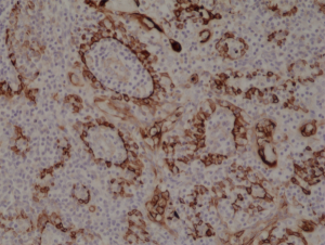
Immunohistochemical staining of formalin fixed and paraffin embedded human tonsil tissue section using anti-CK-17 rabbit monoclonal antibody (Clone RM351) at a 1:1000 dilution.
Immunohistochemical staining of formalin fixed and paraffin embedded human tonsil tissue section using anti-CK-17 rabbit monoclonal antibody (Clone RM351) at a 1:1000 dilution.
anti-Cytokeratin-17 (human), Rabbit Monoclonal (RM351)
REV-31-1237-00
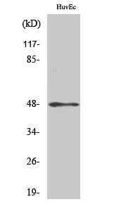
ApplicationsWestern Blot, ImmunoHistoChemistry
Product group Antibodies
ReactivityHuman
TargetKRT17
Overview
- SupplierRevMAb Biosciences
- Product Nameanti-Cytokeratin-17 (human), Rabbit Monoclonal (RM351)
- Delivery Days Customer2
- ApplicationsWestern Blot, ImmunoHistoChemistry
- CertificationResearch Use Only
- ClonalityMonoclonal
- Clone IDRM351
- Gene ID3872
- Target nameKRT17
- Target descriptionkeratin 17
- Target synonyms39.1, CK-17, K17, PC, PC2, PCHC1, keratin, type I cytoskeletal 17, cytokeratin-17, keratin 17, type I
- HostRabbit
- IsotypeIgG
- Protein IDQ04695
- Protein NameKeratin, type I cytoskeletal 17
- Scientific DescriptionCytokeratins are keratin proteins found in the intracytoplasmic cytoskeleton of epithelial tissue (at least 20 different polypeptides). They are an important component of intermediate filaments, which help cells resist mechanical stress. Expression of these cytokeratins within epithelial cells is largely specific to particular organs or tissues. The subsets of cytokeratins which an epithelial cell expresses depends mainly on the type of epithelium, the moment in the course of terminal differentiation and the stage of development. Thus a specific cytokeratin expression profile allows the identification of epithelial cells. Furthermore, this applies also to the malignant counterparts of the epithelia, (carcinomas). Cytokeratin subtype expression patterns are used to an increasing extent in the distinction of different types of epithelial malignancies. The cytokeratin antibodies are not only of assistance in the differential diagnosis of tumors using immunohistochemistry on tissue sections, but are also a useful tool in cytopathology and flow cytometric assays. Cytokeratin-17 is a type I cytokeratin. It is found in nail beds, hair follicles, sebaceous glands and other epidermal appendages. Cytokeratin-17 may be used to aid in the identification of squamous cell carcinomas in various tissues including the cervix, lung, and oral cavity. Mutations in the gene encoding this protein lead to PC-K17 (previously known as Jackson-Lawler) type pachyonychia congenita and steatocystoma multiplex. - Recombinant Antibody. This antibody reacts to human Cytokeratin 17 (CK-17). Applications: WB, IHC. Source: Rabbit. Liquid. 50% Glycerol/PBS with 1% BSA and 0.09% sodium azide. Cytokeratins are keratin proteins found in the intracytoplasmic cytoskeleton of epithelial tissue (at least 20 different polypeptides). They are an important component of intermediate filaments, which help cells resist mechanical stress. Expression of these cytokeratins within epithelial cells is largely specific to particular organs or tissues. The subsets of cytokeratins which an epithelial cell expresses depends mainly on the type of epithelium, the moment in the course of terminal differentiation and the stage of development. Thus a specific cytokeratin expression profile allows the identification of epithelial cells. Furthermore, this applies also to the malignant counterparts of the epithelia, (carcinomas). Cytokeratin subtype expression patterns are used to an increasing extent in the distinction of different types of epithelial malignancies. The cytokeratin antibodies are not only of assistance in the differential diagnosis of tumors using immunohistochemistry on tissue sections, but are also a useful tool in cytopathology and flow cytometric assays. Cytokeratin-17 is a type I cytokeratin. It is found in nail beds, hair follicles, sebaceous glands and other epidermal appendages. Cytokeratin-17 may be used to aid in the identification of squamous cell carcinomas in various tissues including the cervix, lung, and oral cavity. Mutations in the gene encoding this protein lead to PC-K17 (previously known as Jackson-Lawler) type pachyonychia congenita and steatocystoma multiplex.
- ReactivityHuman
- Storage Instruction-20°C,2°C to 8°C
- UNSPSC41116161
Datasheet
Related products
Product group Antibodies

Anti-Keratin 17 [A4]AB00405-1.1-BT
ApplicationsWestern Blot, ELISA, ImmunoHistoChemistry
ReactivityHuman
TargetKRT17
- SizePrice
Product group Antibodies
ApplicationsImmunoFluorescence, Western Blot, ELISA, ImmunoHistoChemistry
ReactivityHuman, Mouse, Rat
- SizePrice

Product group Antibodies
ApplicationsFlow Cytometry, ImmunoFluorescence, Western Blot, ImmunoCytoChemistry, ImmunoHistoChemistry
ReactivityHuman, Mouse, Rat
TargetKRT17
- SizePrice

Product group Antibodies
Anti-KRT17 AntibodyHPA000452
ApplicationsWestern Blot, ImmunoCytoChemistry, ImmunoHistoChemistry
ReactivityHuman, Rat
TargetKRT17
- SizePrice
Product group Antibodies
KRT17 Monoclonal AntibodyCSB-MA000239
ApplicationsImmunoPrecipitation, Western Blot, ELISA
ReactivityHuman
TargetKRT17
- SizePrice
Product group Antibodies

ApplicationsWestern Blot, ELISA
ReactivityHuman
TargetKRT17
- SizePrice
Product group Antibodies

Mouse anti Cytokeratin 17 / Keratin K17MUB0325P-CE/IVD
ApplicationsFlow Cytometry, Western Blot, ImmunoCytoChemistry, ImmunoHistoChemistry, ImmunoHistoChemistry Frozen, ImmunoHistoChemistry Paraffin
ReactivityHuman, Rat
TargetKRT17
- SizePrice
Product group Antibodies
ApplicationsImmunoPrecipitation, Western Blot, ImmunoCytoChemistry, ImmunoHistoChemistry
ReactivityMouse, Porcine, Rat
TargetKRT17
- SizePrice

Product group Antibodies
Cytokeratin 17 Recombinant AntibodyBSM-52057R
ApplicationsFlow Cytometry, ImmunoFluorescence, Western Blot, ImmunoCytoChemistry, ImmunoHistoChemistry, ImmunoHistoChemistry Frozen, ImmunoHistoChemistry Paraffin
ReactivityHuman, Mouse, Rat
TargetKRT17
- SizePrice