

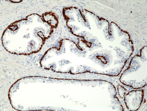
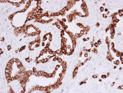
Immunohistochemical staining of formalin fixed and paraffin embedded human Prostate tissue section using anti-CK-5/CK-6 rabbit monoclonal antibody (Clone RM341) at a 1:1000 dilution.
Immunohistochemical staining of formalin fixed and paraffin embedded human Prostate tissue section using anti-CK-5/CK-6 rabbit monoclonal antibody (Clone RM341) at a 1:1000 dilution.
anti-Cytokeratin-5 / -6 (human), Rabbit Monoclonal (RM341)
REV-31-1227-00
ApplicationsWestern Blot, ImmunoHistoChemistry
Product group Antibodies
ReactivityHuman
TargetKRT5
Overview
- SupplierRevMAb Biosciences
- Product Nameanti-Cytokeratin-5 / -6 (human), Rabbit Monoclonal (RM341)
- Delivery Days Customer2
- ApplicationsWestern Blot, ImmunoHistoChemistry
- CertificationResearch Use Only
- ClonalityMonoclonal
- Clone IDRM341
- Gene ID3852
- Target nameKRT5
- Target descriptionkeratin 5
- Target synonymsCK5, DDD, DDD1, EBS1, EBS2, EBS2A, EBS2B, EBS2C, EBS2D, EBS2E, EBS2F, K5, KRT5A, keratin, type II cytoskeletal 5, 58 kda cytokeratin, CK-5, cytokeratin-5, epidermolysis bullosa simplex 2 Dowling-Meara/Kobner/Weber-Cockayne types, keratin 5 (epidermolysis bullosa simplex, Dowling-Meara/Kobner/Weber-Cockayne types), keratin 5, type II, type-II keratin Kb5
- HostRabbit
- IsotypeIgG
- Protein IDP13647
- Protein NameKeratin, type II cytoskeletal 5
- Scientific DescriptionCytokeratins are keratin proteins found in the intracytoplasmic cytoskeleton of epithelial tissue (at least 20 different polypeptides). They are an important component of intermediate filaments, which help cells resist mechanical stress. Expression of these cytokeratins within epithelial cells is largely specific to particular organs or tissues. The subsets of cytokeratins which an epithelial cell expresses depends mainly on the type of epithelium, the moment in the course of terminal differentiation and the stage of development. Thus a specific cytokeratin expression profile allows the identification of epithelial cells. Furthermore, this applies also to the malignant counterparts of the epithelia, (carcinomas). Cytokeratin subtype expression patterns are used to an increasing extent in the distinction of different types of epithelial malignancies. The cytokeratin antibodies are not only of assistance in the differential diagnosis of tumors using immunohistochemistry on tissue sections, but are also a useful tool in cytopathology and flow cytometric assays. Cytokeratin-5/-6 are type II cytokeratins specifically expressed in the basal layer of the epidermis. Targeting both cytokeratin 5 and cytokeratin 6 is used in immunohistochemistry. This often called CK5/6 staining is used for identifying basal cells or myoepithelial cells in the breast and prostate, for breast pathology in distinguishing usual ductal hyperplasia (UDH) and papillary lesions from ductal carcinoma in situ, to distinguish between the diagnosis of papilloma or papillary carcinoma, and in the lung, distinguishing epithelioid mesothelioma (CK5/6 positive in 83%) from lung adenocarcinoma (CK5/6 negative in 85%). - Recombinant Antibody. This antibody reacts to both human CK-5 (Cytokeratin 5) and CK-6 (Cytokeratin 6). Applications: WB, IHC. Source: Rabbit. Liquid. 50% Glycerol/PBS with 1% BSA and 0.09% sodium azide. Cytokeratins are keratin proteins found in the intracytoplasmic cytoskeleton of epithelial tissue (at least 20 different polypeptides). They are an important component of intermediate filaments, which help cells resist mechanical stress. Expression of these cytokeratins within epithelial cells is largely specific to particular organs or tissues. The subsets of cytokeratins which an epithelial cell expresses depends mainly on the type of epithelium, the moment in the course of terminal differentiation and the stage of development. Thus a specific cytokeratin expression profile allows the identification of epithelial cells. Furthermore, this applies also to the malignant counterparts of the epithelia, (carcinomas). Cytokeratin subtype expression patterns are used to an increasing extent in the distinction of different types of epithelial malignancies. The cytokeratin antibodies are not only of assistance in the differential diagnosis of tumors using immunohistochemistry on tissue sections, but are also a useful tool in cytopathology and flow cytometric assays. Cytokeratin-5/-6 are type II cytokeratins specifically expressed in the basal layer of the epidermis. Targeting both cytokeratin 5 and cytokeratin 6 is used in immunohistochemistry. This often called CK5/6 staining is used for identifying basal cells or myoepithelial cells in the breast and prostate, for breast pathology in distinguishing usual ductal hyperplasia (UDH) and papillary lesions from ductal carcinoma in situ, to distinguish between the diagnosis of papilloma or papillary carcinoma, and in the lung, distinguishing epithelioid mesothelioma (CK5/6 positive in 83%) from lung adenocarcinoma (CK5/6 negative in 85%).
- ReactivityHuman
- Storage Instruction-20°C,2°C to 8°C
- UNSPSC41116161
Datasheet
Related products
Product group Antibodies

Anti-Basal cell Cytokeratin [RCK103]AB03334-1.1
ApplicationsFlow Cytometry, ImmunoHistoChemistry
ReactivityAvian, Canine, Chicken, Guinea Pig, Hamster, Human, Porcine, Rabbit, Rat, Zebra Fish
TargetKRT5
- SizePrice
Product group Antibodies
Anti-KRT5 Antibody144-02662
ApplicationsImmunoFluorescence, Western Blot, ImmunoHistoChemistry
ReactivityHuman, Mouse
TargetKRT5
- SizePrice

Product group Antibodies
Anti-KRT5 AntibodyAMAB91549
ApplicationsWestern Blot, ImmunoCytoChemistry, ImmunoHistoChemistry
ReactivityHuman
TargetKRT5
- SizePrice
Product group Antibodies
ApplicationsImmunoFluorescence, Western Blot, ELISA, ImmunoHistoChemistry
ReactivityHuman, Mouse, Rat
- SizePrice
Product group Antibodies

KRT5 / CK5 / Cytokeratin 5 AntibodyLS-C835149
ApplicationsImmunoHistoChemistry
ReactivityHuman
TargetKRT5
- SizePrice

Product group Antibodies
Anti-Cytokeratin 5/KRT5 Antibody Picoband(r)A00398-CARRIER-FREE
ApplicationsWestern Blot, ImmunoHistoChemistry
ReactivityHuman
TargetKRT5
- SizePrice
Product group Antibodies
ApplicationsWestern Blot
ReactivityBovine, Canine, Equine, Human, Mouse, Porcine, Rabbit, Rat
TargetKRT5
- SizePrice
Product group Antibodies
KRT5 AntibodyCSB-PA002049
ApplicationsImmunoFluorescence, Western Blot, ELISA, ImmunoHistoChemistry
ReactivityHuman, Mouse, Rat
TargetKRT5
- SizePrice
Product group Antibodies
Cytokeratin 5 antibodyGTX113219
ApplicationsImmunoFluorescence, Western Blot, ImmunoCytoChemistry, ImmunoHistoChemistry, ImmunoHistoChemistry Paraffin
ReactivityHuman, Mouse
TargetKRT5
- SizePrice