

Anti-EpCAM [3-17I]
AB02662-3.0-BT
ApplicationsFlow Cytometry, ELISA, ImmunoHistoChemistry
Product group Antibodies
ReactivityHuman, Monkey
TargetEPCAM
Overview
- SupplierAbsolute Antibody
- Product NameAnti-EpCAM [3-17I]
- Delivery Days Customer9
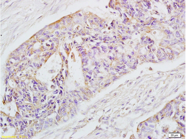
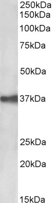
- Application Supplier NoteThe antibody specifically interacts with the natural EpCAM+Kato III cell line by flow cytometric analysis. The IgG format of the antibody bound to both human and cynomolgus EpCAM under non-reducing conditions in western blot analysis. In contrast, no binding of the antibody was observed to any reduced EpCAM antigen. Further, ELISA data demonstrated good binding of the scFv and IgG formats of the antibody to both human and cynomolgus EpCAM. The binding affinity of the IgG1 format to human and monkey EpCAM was measured by surface plasmon resonance (Kd= 1 nM and 0.93 nM, respectively). The ability of the IgG format of the antibody to induce ADCC was analyzed using three different breast cancer cell lines: MDA-MB-231, MDA-453, and BT-474. The results clearly demonstrated the antibody-induced ADCC in all three cell lines, MDA-MB-453, MDA-MB-231, and BT-474, in the presence of human PBMCs with respective estimated EC50 values of 0.08 ng/ml, 15 ng/ml, and 0.12 ng/ml. The maximum killings achieved were 75%, 92%, and 61%, respectively. The ability of the IgG version of the antibody to induce complement-dependent cytotoxicity (CDC) was analyzed using two cell lines, KATO III and MT-3. The results clearly demonstrated that the antibody-induced CDC in the cell lines KATO III and MT-3 in the presence of human serum (EC50 = 0.28 ng/ml and 0.38 ng/ml for KATO III and MT-3 cells, respectively) (US8637017B2). Immunohistochemistry studies of the reactivity of the IgG2A format of the antibody were performed on a panel of normal human tissues. The antibody showed a strong reaction to sloughed cells of the esophagus sample, colon, breast, and lung cancers with epithelial origin. The antibody was conjugated to Strepsaporin. PCI of 3-17I-saporin attenuated cellular viability in three different EpCAM-positive cancer cell lines (MCF7, BxPC-3, and WiDr) as measured by MTS assay. Further, PCI of 3-17I-saporin attenuated both the proliferation and colony-forming ability of EpCAM-positive MCF7 cells (Lund et al., 2014; PMID: 24525727).
- ApplicationsFlow Cytometry, ELISA, ImmunoHistoChemistry
- CertificationResearch Use Only
- ClonalityMonoclonal
- Clone ID3-17I
- Gene ID4072
- Target nameEPCAM
- Target descriptionepithelial cell adhesion molecule
- Target synonymsBer-Ep4, BerEp4, DIAR5, EGP-2, EGP314, EGP40, ESA, HNPCC8, KS1/4, KSA, LYNCH8, M4S1, MIC18, MK-1, MOC-31, TACSTD1, TROP1, epithelial cell adhesion molecule, adenocarcinoma-associated antigen, cell surface glycoprotein Trop-1, epithelial glycoprotein 314, human epithelial glycoprotein-2, major gastrointestinal tumor-associated protein GA733-2, membrane component, chromosome 4, surface marker (35kD glycoprotein), trophoblast cell surface antigen 1, tumor-associated calcium signal transducer 1
- HostMouse
- IsotypeIgG2b
- Protein IDP16422
- Protein NameEpithelial cell adhesion molecule
- Scientific DescriptionThis chimeric mouse antibody was made using the variable domain sequences of the original Human IgG1 format, for improved compatibility with existing reagents, assays and techniques.
- ReactivityHuman, Monkey
- Storage Instruction-20°C,2°C to 8°C
- UNSPSC12352203
Related products
Product group Antibodies
Epcam Polyclonal AntibodyCAC11319
ApplicationsImmunoFluorescence, Western Blot, ELISA, ImmunoHistoChemistry
ReactivityRat
TargetEPCAM
- SizePrice
Product group Antibodies
References
EpCAM Polyclonal AntibodyBS-1513R
ApplicationsFlow Cytometry, ImmunoFluorescence, Western Blot, ELISA, ImmunoCytoChemistry, ImmunoHistoChemistry, ImmunoHistoChemistry Frozen, ImmunoHistoChemistry Paraffin
ReactivityHuman, Mouse, Rat
TargetEPCAM
- SizePrice
Product group Antibodies
Anti-EpCAM AntibodyA85204
ApplicationsWestern Blot, ELISA
ReactivityHuman
- SizePrice
Product group Antibodies
Anti-EPCAM Antibody144-01177
ApplicationsWestern Blot, ImmunoHistoChemistry
ReactivityHuman, Mouse
TargetEPCAM
- SizePrice

Product group Antibodies
Anti-EPCAM AntibodyAMAB91411
ApplicationsWestern Blot, ImmunoHistoChemistry
ReactivityHuman
TargetEPCAM
- SizePrice
Product group Antibodies

Anti-EpCAM [AUA1]Ab00609-1.1
ApplicationsFlow Cytometry, ImmunoFluorescence, ELISA, ImmunoHistoChemistry
ReactivityHuman
TargetEPCAM
- SizePrice
Product group Antibodies

ApplicationsWestern Blot, ELISA
ReactivityHuman
TargetEPCAM
- SizePrice
Product group Antibodies
References
EpCAM antibody [N3C3]GTX113091
ApplicationsImmunoFluorescence, Western Blot, ImmunoCytoChemistry, ImmunoHistoChemistry, ImmunoHistoChemistry Paraffin
ReactivityHuman, Mouse
TargetEPCAM
- SizePrice