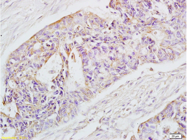
Anti-EpCAM [G8.8]
AB02719-3.3-BT
ApplicationsFlow Cytometry, ImmunoFluorescence, ImmunoPrecipitation, ImmunoHistoChemistry
Product group Antibodies
ReactivityHuman, Mouse
TargetEPCAM
Overview
- SupplierAbsolute Antibody
- Product NameAnti-EpCAM [G8.8]
- Delivery Days Customer9
- Application Supplier NoteTo research scarring in humans, immunofluorescence was preformed on human skin and scar biopsies using the rat version of this antibody (Jiang et al, 2020; pmid:33159076). To show that PDCs expressing the established surface and cancer stem cell marker EpCAM give rise to HCC in inflamed liver, immunohistochemistry was preformed on mouse liver cells using the rat version of this antibody (Matsumotot et al, 2017; pmid:28951464). To show that major facilitator super family domain containing 2a (Mfsd2a), previously known to maintain blood-brain barrier function, is a periportal zonation marker, immunofluorescence was preformed on liver cells from Mfsd2a-CreER mice using the rat version of this antibody (Pu et al, 2016; pmid:27857132). To research the expression of EpCam, immunohistochemistry was preformed on mosue thymus cells using the rat version of this antibody. Further, the antigen of this antibody was immunoprecipitated using the rat version of this antibody bound to sepharose beads (Farr et al, 1991; pmid:2016514). Epithelial cell adhesion molecule expression in A2C12, A549, and Caco-2 cells was determined by flow cytometry using the rat version of this antibody. Furthermore, cell proliferation of A2C12, A549, and Caco-2 cells under the influence of the rat version of this antibody was determined. A significant increase in cell proliferation was observed (Maaser and Borlak, 2008; pmid:19002182). To look at the expression patterns of EpCam., immunohistochemistry was preformed on both human and murine tissue using this antibody. The tissues used were pancreas, prostate, brain and endothelium tissue (Amann et al, 2008; pmid:18172306). To study the expression of EpCam at an early stage of liver development immunohistochemistry was preformed on whole mice embryos and fetal liver using the rat version of this antibody (Tanaka et al, 2009; pmid:19527784). To establish a system to purify spermatogonial stem cells, the rat version of this antibody was used for flow cytometry on testis cells from 5- to 10 week old male mice (Kanatsu-Shinohara et al, 2011; pmid:21858196).
- ApplicationsFlow Cytometry, ImmunoFluorescence, ImmunoPrecipitation, ImmunoHistoChemistry
- CertificationResearch Use Only
- ClonalityMonoclonal
- Clone IDG8.8
- Gene ID4072
- Target nameEPCAM
- Target descriptionepithelial cell adhesion molecule
- Target synonymsBer-Ep4, BerEp4, DIAR5, EGP-2, EGP314, EGP40, ESA, HNPCC8, KS1/4, KSA, LYNCH8, M4S1, MIC18, MK-1, MOC-31, TACSTD1, TROP1, epithelial cell adhesion molecule, adenocarcinoma-associated antigen, cell surface glycoprotein Trop-1, epithelial glycoprotein 314, human epithelial glycoprotein-2, major gastrointestinal tumor-associated protein GA733-2, membrane component, chromosome 4, surface marker (35kD glycoprotein), trophoblast cell surface antigen 1, tumor-associated calcium signal transducer 1
- HostMouse
- IsotypeIgG2b
- Protein IDP16422
- Protein NameEpithelial cell adhesion molecule
- Scientific DescriptionThis chimeric mouse antibody was made using the variable domain sequences of the original Rat IgG2a format, for improved compatibility with existing reagents, assays and techniques.
- ReactivityHuman, Mouse
- Storage Instruction-20°C,2°C to 8°C
- UNSPSC41116161


