Anti-Epiregulin [9E5]
AB03085-10.3-BT
ApplicationsFlow Cytometry, ImmunoFluorescence, Western Blot, Neutralisation/Blocking, Other Application
Product group Antibodies
ReactivityHuman
TargetEREG
Overview
- SupplierAbsolute Antibody
- Product NameAnti-Epiregulin [9E5]
- Delivery Days Customer9
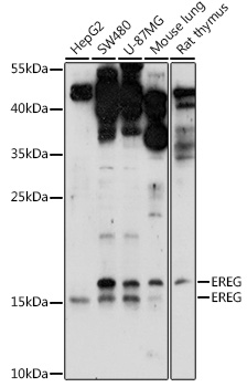
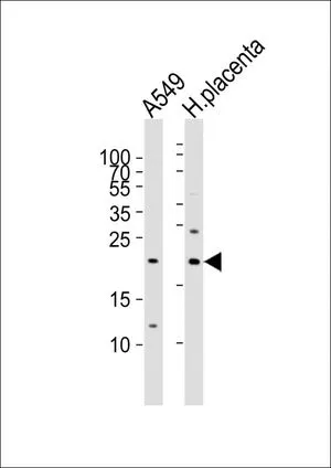
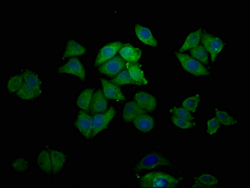
- Application Supplier NoteThe antibody (IgG) and two humanized versions were used to examine the cell surface and subcellular localization of epiregulin by immunofluorescence staining. The antibody-binding assays were performed using colorectal carcinoma cells (DLD-1, HCT116). The original antibody and the humanized version HM1 were detected predominantly as intense green florescence spots in the cell surface with some faint fluorescence demonstrating subcellular distribution in colon cancer cells. The humanized version HM0 also exhibited weak and diffuse cell surface stained. The cell surface binding efficiency of each antibody to epiregulin expressing cells was determined by flow cytometry on DLD-1 and HCT116 cells. The results of flow cytometry indicated that HM1 bound more efficiently to DLD-1 and HCT116 cells than the same concentration of HM0. Further, HM1 was able to bind epiregulin expressing cells at a relatively low concentration (0.1 ug/mL). Surface plasmon resonance assay was performed to evaluate the binding specificity and affinity between the antibody and the humanized versions of the antibody and epiregulin. The antibody was not effective in mediating ADCC in either DLD-1 or HCT116 cells, while the humanized versions showed improved levels of cellular cytotoxicity. (Hun Lee et al, 2013; PMID: 24239549). The crystal structure of the Fab fragment in the presence and absence of EPR was determined (Kado et al., 2016; PMID: 26627827). The antibody efficiently inhibited the induction of EGFR signaling by EREG. The antibody had little effect on cell growth, but was found to inhibit focal adhesion formation and cell spreading of colon cancer cell lines. The antibody detected epiregulin from DLD-1 cell lysates by western blot analysis (Iijima et al., 2017 PMID: 28274874). The antibody was used to develop a nanomedicine composed of a phase-change nano-droplet (PCND) coated with the antibody. The antibody conjugated PCNDs were selectively internalised into targeted cancer cells and kill the cells dynamically by ultrasound-induced intracellular vaporisation. In vitro experiments showed that antibody-conjugated PCND targets 97.8% of high-EREG-expressing cancer cells and kills 57% of those targeted upon exposure to ultrasound. Furthermore, direct observation of the intracellular vaporisation process revealed the significant morphological alterations of cells and the release of intracellular contents (Ishijima et al, 2017).
- ApplicationsFlow Cytometry, ImmunoFluorescence, Western Blot, Neutralisation/Blocking, Other Application
- CertificationResearch Use Only
- ClonalityMonoclonal
- Clone ID9E5
- Gene ID2069
- Target nameEREG
- Target descriptionepiregulin
- Target synonymsEPR, ER, Ep, proepiregulin
- HostHuman
- IsotypeIgG1
- Protein IDO14944
- Protein NameProepiregulin
- Scientific DescriptionThis chimeric human antibody was made using the variable domain sequences of the original Mouse IgG format for improved compatibility with existing reagents assays and techniques.
- ReactivityHuman
- Storage Instruction-20°C,2°C to 8°C
- UNSPSC12352203


