

Figure 1. Western blot analysis of FADD using anti-FADD antibody (PA1464). Electrophoresis was performed on a 5-20% SDS-PAGE gel at 70V (Stacking gel) / 90V (Resolving gel) for 2-3 hours. The sample well of each lane was loaded with 30 ug of sample under reducing conditions. Lane 1: human HT1080 whole cell lysates, Lane 2: human A549 whole cell lysates, Lane 3: human Jurkat whole cell lysates, Lane 4: human A431 whole cell lysates, Lane 5: human HepG2 whole cell lysates, Lane 6: human Hela whole cell lysates, Lane 7: human MCF-7 whole cell lysates, Lane 8: human Raji whole cell lysates. After electrophoresis, proteins were transferred to a nitrocellulose membrane at 150 mA for 50-90 minutes. Blocked the membrane with 5% non-fat milk/TBS for 1.5 hour at RT. The membrane was incubated with rabbit anti-FADD antigen affinity purified polyclonal antibody (Catalog # PA1464) at 0.5 microg/mL overnight at 4°C, then washed with TBS-0.1%Tween 3 times with 5 minutes each and probed with a goat anti-rabbit IgG-HRP secondary antibody at a dilution of 1:5000 for 1.5 hour at RT. The signal is developed using an Enhanced Chemiluminescent detection (ECL) kit (Catalog # EK1002) with Tanon 5200 system. A specific band was detected for FADD at approximately 28 kDa. The expected band size for FADD is at 23 kDa.
. Electrophoresis was performed on a 5-20% SDS-PAGE gel at 70V (Stacking gel) / 90V (Resolving gel) for 2-3 hours. The sample well of each lane was loaded with 30 ug of sample under reducing conditions. Lane 1: rat spleen tissue lysates, Lane 2: rat PC-12 whole cell lysates, Lane 3: mouse spleen tissue lysates, Lane 4: mouse kidney tissue lysates, Lane 5: mouse RAW264.7 whole cell lysates. After electrophoresis, proteins were transferred to a nitrocellulose membrane at 150 mA for 50-90 minutes. Blocked the membrane with 5% non-fat milk/TBS for 1.5 hour at RT. The membrane was incubated with rabbit anti-FADD antigen affinity purified polyclonal antibody (Catalog # PA1464) at 0.5 microg/mL overnight at 4°C, then washed with TBS-0.1%Tween 3 times with 5 minutes each and probed with a goat anti-rabbit IgG-HRP secondary antibody at a dilution of 1:5000 for 1.5 hour at RT. The signal is developed using an Enhanced Chemiluminescent detection (ECL) kit (Catalog # EK1002) with Tanon 5200 system. A specific band was detected for FADD at approximately 28 kDa. The expected band size for FADD is at 23 kDa.")
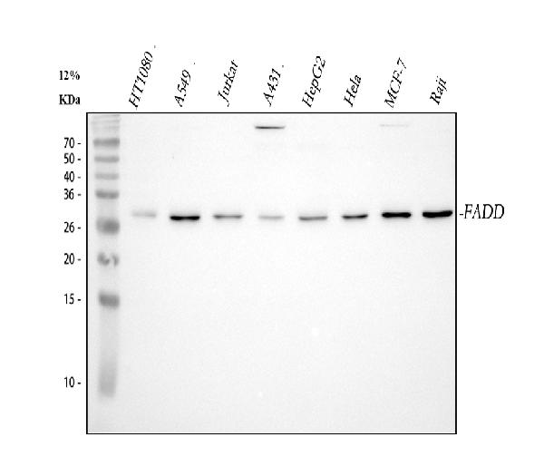
Figure 1. Western blot analysis of FADD using anti-FADD antibody (PA1464). Electrophoresis was performed on a 5-20% SDS-PAGE gel at 70V (Stacking gel) / 90V (Resolving gel) for 2-3 hours. The sample well of each lane was loaded with 30 ug of sample under reducing conditions. Lane 1: human HT1080 whole cell lysates, Lane 2: human A549 whole cell lysates, Lane 3: human Jurkat whole cell lysates, Lane 4: human A431 whole cell lysates, Lane 5: human HepG2 whole cell lysates, Lane 6: human Hela whole cell lysates, Lane 7: human MCF-7 whole cell lysates, Lane 8: human Raji whole cell lysates. After electrophoresis, proteins were transferred to a nitrocellulose membrane at 150 mA for 50-90 minutes. Blocked the membrane with 5% non-fat milk/TBS for 1.5 hour at RT. The membrane was incubated with rabbit anti-FADD antigen affinity purified polyclonal antibody (Catalog # PA1464) at 0.5 microg/mL overnight at 4°C, then washed with TBS-0.1%Tween 3 times with 5 minutes each and probed with a goat anti-rabbit IgG-HRP secondary antibody at a dilution of 1:5000 for 1.5 hour at RT. The signal is developed using an Enhanced Chemiluminescent detection (ECL) kit (Catalog # EK1002) with Tanon 5200 system. A specific band was detected for FADD at approximately 28 kDa. The expected band size for FADD is at 23 kDa.
Anti-FADD Antibody Picoband(r)

PA1464-DYLIGHT550
ApplicationsWestern Blot
Product group Antibodies
ReactivityHuman, Mouse, Rat
TargetFADD
Overview
- SupplierBoster Bio
- Product NameAnti-FADD Antibody Picoband(r)
- Delivery Days Customer9
- Antibody SpecificityNo cross reactivity with other proteins.
- Application Supplier NoteTested Species: In-house tested species with positive results. Predicted Species: Species predicted to be fit for the product based on sequence similarities. Other applications have not been tested. Optimal dilutions should be determined by end users.
- ApplicationsWestern Blot
- CertificationResearch Use Only
- ClonalityPolyclonal
- Concentration500 ug/ml
- ConjugateDyLight 550
- Gene ID8772
- Target nameFADD
- Target descriptionFas associated via death domain
- Target synonymsFas (TNFRSF6)-associated via death domain; FAS-associated death domain protein; Fas-associating death domain-containing protein; Fas-associating protein with death domain; GIG3; growth-inhibiting gene 3 protein; mediator of receptor-induced toxicity; MORT1
- HostRabbit
- IsotypeIgG
- Protein IDQ13158
- Protein NameFAS-associated death domain protein
- Scientific DescriptionBoster Bio Anti-FADD Antibody catalog # PA1464. Tested in WB applications. This antibody reacts with Human, Mouse, Rat. The brand Picoband indicates this is a premium antibody that guarantees superior quality, high affinity, and strong signals with minimal background in Western blot applications. Only our best-performing antibodies are designated as Picoband, ensuring unmatched performance.
- ReactivityHuman, Mouse, Rat
- Storage Instruction-20°C,2°C to 8°C
- UNSPSC12352203