

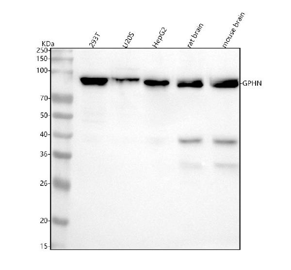
Figure 1. Western blot analysis of Gephyrin using anti-Gephyrin antibody (M04560-3). Electrophoresis was performed on a 5-20% SDS-PAGE gel at 70V (Stacking gel) / 90V (Resolving gel) for 2-3 hours. The sample well of each lane was loaded with 30 ug of sample under reducing conditions. Lane 1: human 293T whole cell lysates, Lane 2: human U20S whole cell lysates, Lane 3: human HepG2 whole cell lysates, Lane 4: rat brain tissue lysates, Lane 5: mouse brain tissue lysates. After electrophoresis, proteins were transferred to a nitrocellulose membrane at 150 mA for 50-90 minutes. Blocked the membrane with 5% non-fat milk/TBS for 1.5 hour at RT. The membrane was incubated with rabbit anti-Gephyrin antigen affinity purified monoclonal antibody (Catalog # M04560-3) at 1:500 overnight at 4°C, then washed with TBS-0.1%Tween 3 times with 5 minutes each and probed with a goat anti-rabbit IgG-HRP secondary antibody at a dilution of 1:5000 for 1.5 hour at RT. The signal is developed using an Enhanced Chemiluminescent detection (ECL) kit (Catalog # EK1002) with Tanon 5200 system. A specific band was detected for Gephyrin at approximately 93 kDa. The expected band size for Gephyrin is at 80 kDa.
Figure 1. Western blot analysis of Gephyrin using anti-Gephyrin antibody (M04560-3). Electrophoresis was performed on a 5-20% SDS-PAGE gel at 70V (Stacking gel) / 90V (Resolving gel) for 2-3 hours. The sample well of each lane was loaded with 30 ug of sample under reducing conditions. Lane 1: human 293T whole cell lysates, Lane 2: human U20S whole cell lysates, Lane 3: human HepG2 whole cell lysates, Lane 4: rat brain tissue lysates, Lane 5: mouse brain tissue lysates. After electrophoresis, proteins were transferred to a nitrocellulose membrane at 150 mA for 50-90 minutes. Blocked the membrane with 5% non-fat milk/TBS for 1.5 hour at RT. The membrane was incubated with rabbit anti-Gephyrin antigen affinity purified monoclonal antibody (Catalog # M04560-3) at 1:500 overnight at 4°C, then washed with TBS-0.1%Tween 3 times with 5 minutes each and probed with a goat anti-rabbit IgG-HRP secondary antibody at a dilution of 1:5000 for 1.5 hour at RT. The signal is developed using an Enhanced Chemiluminescent detection (ECL) kit (Catalog # EK1002) with Tanon 5200 system. A specific band was detected for Gephyrin at approximately 93 kDa. The expected band size for Gephyrin is at 80 kDa.
Anti-Gephyrin Rabbit Monoclonal Antibody
M04560-3
ApplicationsImmunoFluorescence, Western Blot, ImmunoCytoChemistry, ImmunoHistoChemistry
Product group Antibodies
ReactivityHuman, Mouse, Rat
TargetGPHN
Overview
- SupplierBoster Bio
- Product NameAnti-Gephyrin Rabbit Monoclonal Antibody
- Delivery Days Customer9
- ApplicationsImmunoFluorescence, Western Blot, ImmunoCytoChemistry, ImmunoHistoChemistry
- CertificationResearch Use Only
- ClonalityMonoclonal
- Clone ID18G74
- Gene ID10243
- Target nameGPHN
- Target descriptiongephyrin
- Target synonymsGEPH; gephyrin; GPH; GPHRYN; HKPX1; MOCODC
- HostRabbit
- IsotypeIgG
- Protein IDQ9NQX3
- Protein NameGephyrin
- Scientific DescriptionBoster Bio Anti-Gephyrin Rabbit Monoclonal Antibody catalog # M04560-3. Tested in WB, IHC, ICC/IF applications. This antibody reacts with Human, Mouse, Rat.
- ReactivityHuman, Mouse, Rat
- Storage Instruction-20°C
- UNSPSC12352203