

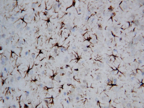
Figure 2. IHC analysis of GFAP using anti-GFAP antibody (PA1239). GFAP was detected in paraffin-embedded section of rat brain tissues. Heat mediated antigen retrieval was performed in citrate buffer (pH6, epitope retrieval solution) for 20 mins. The tissue section was blocked with 10% goat serum. The tissue section was then incubated with 1microg/ml rabbit anti-GFAP Antibody (PA1239) overnight at 4°C. Biotinylated goat anti-rabbit IgG was used as secondary antibody and incubated for 30 minutes at 37°C. The tissue section was developed using Strepavidin-Biotin-Complex (SABC)(Catalog # SA1022) with DAB as the chromogen.
. GFAP was detected in paraffin-embedded section of mouse brain tissues. Heat mediated antigen retrieval was performed in citrate buffer (pH6, epitope retrieval solution ) for 20 mins. The tissue section was blocked with 10% goat serum. The tissue section was then incubated with 1microg/mL rabbit anti-GFAP Antibody (PA1239) overnight at 4°C. Cy3 Conjugated Goat Anti-Rabbit IgG (BA1032) was used as secondary antibody at 1:100 dilution and incubated for 30 minutes at 37°C. The section was counterstained with DAPI. Visualize using a fluorescence microscope and filter sets appropriate for the label used.")
. Electrophoresis was performed on a 5-20% SDS-PAGE gel at 70V (Stacking gel) / 90V (Resolving gel) for 2-3 hours. The sample well of each lane was loaded with 30 ug of sample under reducing conditions. Lane 1: human U251 whole cell lysates, Lane 2: rat brain tissue lysates, Lane 3: mouse brain tissue lysates. After electrophoresis, proteins were transferred to a nitrocellulose membrane at 150 mA for 50-90 minutes. Blocked the membrane with 5% non-fat milk/TBS for 1.5 hour at RT. The membrane was incubated with rabbit anti-GFAP antigen affinity purified polyclonal antibody (Catalog # PA1239) at 0.5 microg/mL overnight at 4°C, then washed with TBS-0.1%Tween 3 times with 5 minutes each and probed with a goat anti-rabbit IgG-HRP secondary antibody at a dilution of 1:5000 for 1.5 hour at RT. The signal is developed using an Enhanced Chemiluminescent detection (ECL) kit (Catalog # EK1002) with Tanon 5200 system. A specific band was detected for GFAP at approximately 50 kDa. The expected band size for GFAP is at 50 kDa.")
. GFAP was detected in a spinal cord. Heat mediated antigen retrieval was performed in EDTA buffer (pH 8.0, epitope retrieval solution). The tissue section was blocked with 10% horse serum. The tissue section was then incubated with anti-GFAP antibody (PA1239) at 1:500 overnight at 4°C. Peroxidase Conjugated Goat Anti-rabbit IgG was used as secondary antibody and incubated for 30 minutes at 37°C. The tissue section was developed using confocal microscope.")
Figure 2. IHC analysis of GFAP using anti-GFAP antibody (PA1239). GFAP was detected in paraffin-embedded section of rat brain tissues. Heat mediated antigen retrieval was performed in citrate buffer (pH6, epitope retrieval solution) for 20 mins. The tissue section was blocked with 10% goat serum. The tissue section was then incubated with 1microg/ml rabbit anti-GFAP Antibody (PA1239) overnight at 4°C. Biotinylated goat anti-rabbit IgG was used as secondary antibody and incubated for 30 minutes at 37°C. The tissue section was developed using Strepavidin-Biotin-Complex (SABC)(Catalog # SA1022) with DAB as the chromogen.
Anti-GFAP Antibody Picoband(r)

PA1239
ApplicationsImmunoFluorescence, Western Blot, ImmunoHistoChemistry
Product group Antibodies
ReactivityChicken, Human, Mouse, Rat
TargetGFAP
Overview
- SupplierBoster Bio
- Product NameAnti-GFAP Antibody
- Delivery Days Customer9
- Antibody SpecificityNo cross reactivity with other proteins.
- Application Supplier NoteTested Species: In-house tested species with positive results. Predicted Species: Species predicted to be fit for the product based on sequence similarities. By Heat: Boiling the paraffin sections in 10mM citrate buffer, pH6.0, for 20mins is required for the staining of formalin/paraffin sections. Other applications have not been tested. Optimal dilutions should be determined by end users.
- ApplicationsImmunoFluorescence, Western Blot, ImmunoHistoChemistry
- Applications SupplierIHP, WB, IHC
- CertificationResearch Use Only
- ClonalityPolyclonal
- Concentration500 ug/ml
- FormulationLyophilized
- Gene ID2670
- Target nameGFAP
- Target descriptionglial fibrillary acidic protein
- Target synonymsALXDRD; glial fibrillary acidic protein
- HostRabbit
- IsotypeIgG
- Protein IDP14136
- Protein NameGlial fibrillary acidic protein
- Scientific DescriptionBoster Bio Anti-GFAP Antibody catalog # PA1239. Tested in IF, IHC, WB applications. This antibody reacts with Human, Mouse, Rat. The brand Picoband indicates this is a premium antibody that guarantees superior quality, high affinity, and strong signals with minimal background in Western blot applications. Only our best-performing antibodies are designated as Picoband, ensuring unmatched performance.
- ReactivityChicken, Human, Mouse, Rat
- Reactivity SupplierHuman, Mouse, Rat, Chicken
- Storage Instruction-20°C,2°C to 8°C
- UNSPSC12352203
References
- CXCR4 knockout induces neuropathological changes in the MPTP-lesioned model of Parkinsons disease. Ma J et al., 2023 Feb, Biochim Biophys Acta Mol Basis DisRead more
- Abnormal Glu/mGluR(2/3)/PI3K pathway in the hippocampal neurovascular unit leads to diabetes-related depression. Liu J et al., 2021 Apr, Neural Regen ResRead more
- OLIG2 Drives Abnormal Neurodevelopmental Phenotypes in Human iPSC-Based Organoid and Chimeric Mouse Models of Down Syndrome. Xu R et al., 2019 Jun 6, Cell Stem CellRead more
- Inhibitory effects of glucagon-like peptide-1 receptor on epilepsy. Wen Y et al., 2019 Mar 26, Biochem Biophys Res CommunRead more
- Neuroprotective effects of the novel GLP-1 long acting analogue semaglutide in the MPTP Parkinsons disease mouse model. Zhang L et al., 2018 Oct, NeuropeptidesRead more
- UCH-L1 inhibition aggravates mossy fiber sprouting in the pentylenetetrazole kindling model. Wen Y et al., 2018 Sep 18, Biochem Biophys Res CommunRead more
- Matrine promotes NT3 expression in CNS cells in experimental autoimmune encephalomyelitis. Zhang ML et al., 2017 May 10, Neurosci LettRead more
- Activation of LILRB2 signal pathway in temporal lobe epilepsy patients and in a pilocarpine induced epilepsy model. Yue J et al., 2016 Nov, Exp NeurolRead more
- Expression of Glypican-4 in the brains of epileptic patients and epileptic animals and its effects on epileptic seizures. Xiong Y et al., 2016 Sep 9, Biochem Biophys Res CommunRead more
- Sulfiredoxin-1 protects primary cultured astrocytes from ischemia-induced damage. Yu S et al., 2015 Mar, Neurochem IntRead more