


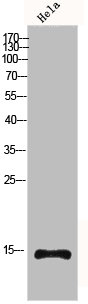
Figure 1. Western blot analysis of Histone H4 using anti-Histone H4 antibody (M14495-2). Electrophoresis was performed on a 5-20% SDS-PAGE gel at 70V (Stacking gel) / 90V (Resolving gel) for 2-3 hours. The sample well of each lane was loaded with 30 ug of sample under reducing conditions. Lane 1: human Hela whole cell lysates, Lane 2: human PC-3 whole cell lysates, Lane 3: human U20S whole cell lysates, Lane 4: human Hacat whole cell lysates, Lane 5: rat brain tissue lysates, Lane 6: rat C6 whole cell lysates, Lane 7: mouse brain tissue lysates, Lane 8: mouse 3T3-L1 whole cell lysates. After electrophoresis, proteins were transferred to a nitrocellulose membrane at 150 mA for 50-90 minutes. Blocked the membrane with 5% non-fat milk/TBS for 1.5 hour at RT. The membrane was incubated with rabbit anti-Histone H4 antigen affinity purified monoclonal antibody (Catalog # M14495-2) at 1:500 overnight at 4°C, then washed with TBS-0.1%Tween 3 times with 5 minutes each and probed with a goat anti-rabbit IgG-HRP secondary antibody at a dilution of 1:1000 for 1.5 hour at RT. The signal is developed using an Enhanced Chemiluminescent detection (ECL) kit (Catalog # EK1002) with Tanon 5200 system. A specific band was detected for Histone H4 at approximately 11 kDa. The expected band size for Histone H4 is at 11 kDa.
. Histone H4 was detected in a paraffin-embedded section of human breast cancer tissue. Heat mediated antigen retrieval was performed in EDTA buffer (pH 8.0, epitope retrieval solution). The tissue section was blocked with 10% goat serum. The tissue section was then incubated with 1:100 rabbit anti-Histone H4 Antibody (M14495-2) overnight at 4°C. Peroxidase Conjugated Goat Anti-rabbit IgG was used as secondary antibody and incubated for 30 minutes at 37°C. The tissue section was developed using HRP Conjugated Rabbit IgG Super Vision Assay Kit (Catalog # SV0002) with DAB as the chromogen.")
. Histone H4 was detected in a paraffin-embedded section of human colorectal adenocarcinoma tissue. Heat mediated antigen retrieval was performed in EDTA buffer (pH 8.0, epitope retrieval solution). The tissue section was blocked with 10% goat serum. The tissue section was then incubated with 1:100 rabbit anti-Histone H4 Antibody (M14495-2) overnight at 4°C. Peroxidase Conjugated Goat Anti-rabbit IgG was used as secondary antibody and incubated for 30 minutes at 37°C. The tissue section was developed using HRP Conjugated Rabbit IgG Super Vision Assay Kit (Catalog # SV0002) with DAB as the chromogen.")
. Histone H4 was detected in a paraffin-embedded section of human liver cancer tissue. Heat mediated antigen retrieval was performed in EDTA buffer (pH 8.0, epitope retrieval solution). The tissue section was blocked with 10% goat serum. The tissue section was then incubated with 1:100 rabbit anti-Histone H4 Antibody (M14495-2) overnight at 4°C. Peroxidase Conjugated Goat Anti-rabbit IgG was used as secondary antibody and incubated for 30 minutes at 37°C. The tissue section was developed using HRP Conjugated Rabbit IgG Super Vision Assay Kit (Catalog # SV0002) with DAB as the chromogen.")
. Histone H4 was detected in a paraffin-embedded section of human lung squamous cell carcinoma tissue. Heat mediated antigen retrieval was performed in EDTA buffer (pH 8.0, epitope retrieval solution). The tissue section was blocked with 10% goat serum. The tissue section was then incubated with 1:100 rabbit anti-Histone H4 Antibody (M14495-2) overnight at 4°C. Peroxidase Conjugated Goat Anti-rabbit IgG was used as secondary antibody and incubated for 30 minutes at 37°C. The tissue section was developed using HRP Conjugated Rabbit IgG Super Vision Assay Kit (Catalog # SV0002) with DAB as the chromogen.")
. Histone H4 was detected in a paraffin-embedded section of human testicular germ cell tumor tissue. Heat mediated antigen retrieval was performed in EDTA buffer (pH 8.0, epitope retrieval solution). The tissue section was blocked with 10% goat serum. The tissue section was then incubated with 1:100 rabbit anti-Histone H4 Antibody (M14495-2) overnight at 4°C. Peroxidase Conjugated Goat Anti-rabbit IgG was used as secondary antibody and incubated for 30 minutes at 37°C. The tissue section was developed using HRP Conjugated Rabbit IgG Super Vision Assay Kit (Catalog # SV0002) with DAB as the chromogen.")
. Histone H4 was detected in a paraffin-embedded section of human placenta tissue. Heat mediated antigen retrieval was performed in EDTA buffer (pH 8.0, epitope retrieval solution). The tissue section was blocked with 10% goat serum. The tissue section was then incubated with 1:100 rabbit anti-Histone H4 Antibody (M14495-2) overnight at 4°C. Peroxidase Conjugated Goat Anti-rabbit IgG was used as secondary antibody and incubated for 30 minutes at 37°C. The tissue section was developed using HRP Conjugated Rabbit IgG Super Vision Assay Kit (Catalog # SV0002) with DAB as the chromogen.")
. Histone H4 was detected in a paraffin-embedded section of human thyroid cancer tissue. Heat mediated antigen retrieval was performed in EDTA buffer (pH 8.0, epitope retrieval solution). The tissue section was blocked with 10% goat serum. The tissue section was then incubated with 1:100 rabbit anti-Histone H4 Antibody (M14495-2) overnight at 4°C. Peroxidase Conjugated Goat Anti-rabbit IgG was used as secondary antibody and incubated for 30 minutes at 37°C. The tissue section was developed using HRP Conjugated Rabbit IgG Super Vision Assay Kit (Catalog # SV0002) with DAB as the chromogen.")
. Histone H4 was detected in a paraffin-embedded section of mouse brain tissue. Heat mediated antigen retrieval was performed in EDTA buffer (pH 8.0, epitope retrieval solution). The tissue section was blocked with 10% goat serum. The tissue section was then incubated with 1:100 rabbit anti-Histone H4 Antibody (M14495-2) overnight at 4°C. Peroxidase Conjugated Goat Anti-rabbit IgG was used as secondary antibody and incubated for 30 minutes at 37°C. The tissue section was developed using HRP Conjugated Rabbit IgG Super Vision Assay Kit (Catalog # SV0002) with DAB as the chromogen.")
. Histone H4 was detected in a paraffin-embedded section of rat brain tissue. Heat mediated antigen retrieval was performed in EDTA buffer (pH 8.0, epitope retrieval solution). The tissue section was blocked with 10% goat serum. The tissue section was then incubated with 1:100 rabbit anti-Histone H4 Antibody (M14495-2) overnight at 4°C. Peroxidase Conjugated Goat Anti-rabbit IgG was used as secondary antibody and incubated for 30 minutes at 37°C. The tissue section was developed using HRP Conjugated Rabbit IgG Super Vision Assay Kit (Catalog # SV0002) with DAB as the chromogen.")
Figure 1. Western blot analysis of Histone H4 using anti-Histone H4 antibody (M14495-2). Electrophoresis was performed on a 5-20% SDS-PAGE gel at 70V (Stacking gel) / 90V (Resolving gel) for 2-3 hours. The sample well of each lane was loaded with 30 ug of sample under reducing conditions. Lane 1: human Hela whole cell lysates, Lane 2: human PC-3 whole cell lysates, Lane 3: human U20S whole cell lysates, Lane 4: human Hacat whole cell lysates, Lane 5: rat brain tissue lysates, Lane 6: rat C6 whole cell lysates, Lane 7: mouse brain tissue lysates, Lane 8: mouse 3T3-L1 whole cell lysates. After electrophoresis, proteins were transferred to a nitrocellulose membrane at 150 mA for 50-90 minutes. Blocked the membrane with 5% non-fat milk/TBS for 1.5 hour at RT. The membrane was incubated with rabbit anti-Histone H4 antigen affinity purified monoclonal antibody (Catalog # M14495-2) at 1:500 overnight at 4°C, then washed with TBS-0.1%Tween 3 times with 5 minutes each and probed with a goat anti-rabbit IgG-HRP secondary antibody at a dilution of 1:1000 for 1.5 hour at RT. The signal is developed using an Enhanced Chemiluminescent detection (ECL) kit (Catalog # EK1002) with Tanon 5200 system. A specific band was detected for Histone H4 at approximately 11 kDa. The expected band size for Histone H4 is at 11 kDa.
Anti-Histone H4 HIST1H4A Rabbit Monoclonal Antibody
M14495-2
ApplicationsImmunoFluorescence, Western Blot, ImmunoCytoChemistry, ImmunoHistoChemistry
Product group Antibodies
ReactivityHuman, Mouse, Rat
TargetH4C9
Overview
- SupplierBoster Bio
- Product NameAnti-Histone H4 HIST1H4A Rabbit Monoclonal Antibody
- Delivery Days Customer9
- ApplicationsImmunoFluorescence, Western Blot, ImmunoCytoChemistry, ImmunoHistoChemistry
- CertificationResearch Use Only
- ClonalityMonoclonal
- Clone IDHDC-8
- Gene ID8294
- Target nameH4C9
- Target descriptionH4 clustered histone 9
- Target synonymsH4-16, H4/m, H4C1, H4C11, H4C12, H4C13, H4C14, H4C15, H4C16, H4C2, H4C3, H4C4, H4C5, H4C6, H4C8, H4FM, H4M, HIST1H4I, TEBIVANED4, TEVANED4, histone H4, H4 histone family, member M, Histone 4 family, member M, histone 1, H4i, histone cluster 1 H4 family member i, histone cluster 1, H4i, histone family member
- HostRabbit
- IsotypeIgG
- Protein IDP62805
- Protein NameHistone H4
- Scientific DescriptionBoster Bio Anti-Histone H4 HIST1H4A Rabbit Monoclonal Antibody catalog # M14495-2. Tested in WB, IHC, ICC/IF applications. This antibody reacts with Human, Mouse, Rat.
- ReactivityHuman, Mouse, Rat
- Storage Instruction-20°C
- UNSPSC12352203
Datasheet
MSDS
Related products
Product group Antibodies
Histone H4 AntibodyCSB-PA002932
ApplicationsImmunoFluorescence, Western Blot, ELISA, ImmunoHistoChemistry
ReactivityHuman, Monkey, Mouse, Rat
TargetH4C9
- SizePrice

Product group Antibodies
Anti-Histone H4 AntibodyA94541
ApplicationsImmunoFluorescence, Western Blot, ELISA, ImmunoHistoChemistry
ReactivityHuman, Mouse, Rat
- SizePrice
Product group Antibodies

ApplicationsWestern Blot, ImmunoHistoChemistry
- SizePrice
Product group Antibodies

Anti-Histone H4 [1A9D7]Ab03227-1.1
ApplicationsWestern Blot, ELISA, Other Application
ReactivityHuman
TargetH4C9
- SizePrice

Product group Antibodies
Anti-HIST4H4 AntibodyHPA042201
ApplicationsImmunoHistoChemistry
ReactivityHuman
TargetH4C9
- SizePrice
Product group Antibodies

Histone H4 AntibodyLS-C401737
ApplicationsWestern Blot, ELISA, ImmunoHistoChemistry
ReactivityHuman, Mouse, Rat
- SizePrice
Product group Antibodies
HIST1H4A Polyclonal AntibodyCAC14537
ApplicationsWestern Blot, ELISA, ImmunoHistoChemistry
ReactivityMouse
TargetH4C9
- SizePrice
Product group Antibodies
ApplicationsDot Blot, ImmunoFluorescence, Western Blot, ChIP Chromatin ImmunoPrecipitation, ELISA, ImmunoCytoChemistry
ReactivityHuman, Mouse
TargetH4C9
- SizePrice
Product group Antibodies
ApplicationsWestern Blot, ELISA, ImmunoCytoChemistry, Other Application
ReactivityVertebrate
TargetH4C9
- SizePrice
Product group Antibodies
ApplicationsDot Blot, ChIP Chromatin ImmunoPrecipitation, ELISA
ReactivityHuman
TargetH4C9
- SizePrice