

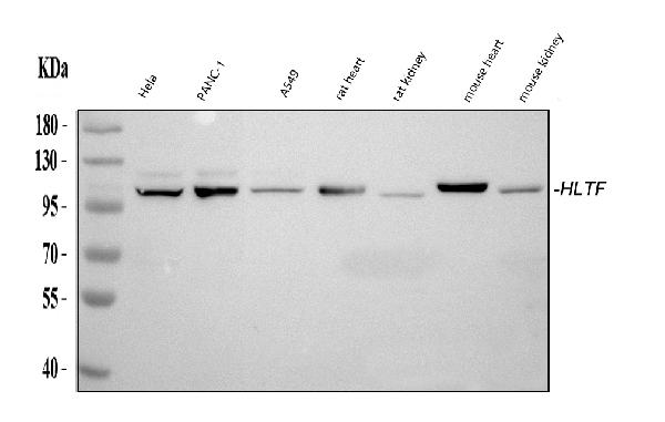
Figure 1. Western blot analysis of HLTF using anti-HLTF antibody (PB10070). Electrophoresis was performed on a 5-20% SDS-PAGE gel at 70V (Stacking gel) / 90V (Resolving gel) for 2-3 hours. The sample well of each lane was loaded with 30 ug of sample under reducing conditions. Lane 1: human Hela whole cell lysates, Lane 2: human PANC-1 whole cell lysates, Lane 3: human A549 whole cell lysates, Lane 4: rat heart tissue lysates, Lane 5: rat kidney tissue lysates, Lane 6: mouse heart tissue lysates, Lane 7: mouse kidney tissue lysates. After electrophoresis, proteins were transferred to a nitrocellulose membrane at 150 mA for 50-90 minutes. Blocked the membrane with 5% non-fat milk/TBS for 1.5 hour at RT. The membrane was incubated with rabbit anti-HLTF antigen affinity purified polyclonal antibody (Catalog # PB10070) at 0.5 microg/mL overnight at 4°C, then washed with TBS-0.1%Tween 3 times with 5 minutes each and probed with a goat anti-rabbit IgG-HRP secondary antibody at a dilution of 1:5000 for 1.5 hour at RT. The signal is developed using an Enhanced Chemiluminescent detection (ECL) kit (Catalog # EK1002) with Tanon 5200 system. A specific band was detected for HLTF at approximately 114 kDa. The expected band size for HLTF is at 114 kDa.
 and anti-Tubulin Alpha antibody (M03989-3). HLTF was detected in immunocytochemical section of A549 cell. Enzyme antigen retrieval was performed using IHC enzyme antigen retrieval reagent (AR0022) for 15 mins. The cells were blocked with 10% goat serum. And then incubated with 5 microg/mL rabbit anti-HLTF Antibody (PB10070) and mouse anti-Tubulin Alpha antibody (M03989-3) overnight at 4°C. Cy3 Conjugated Goat Anti-Rabbit IgG (BA1032) and DyLight®488 Conjugated Goat Anti-Mouse IgG (BA1126) were used as secondary antibody at 1:500 dilution and incubated for 30 minutes at 37°C. Visualize using a fluorescence microscope and filter sets appropriate for the label used.")
. Overlay histogram showing CACO-2 cells stained with PB10070 (Blue line). To facilitate intracellular staining, cells were fixed with 4% paraformaldehyde and permeabilized with permeabilization buffer. The cells were blocked with 10% normal goat serum. And then incubated with rabbit anti-HLTF Antibody (PB10070, 1 microg/1x106 cells) for 30 min at 20°C. DyLight®488 conjugated goat anti-rabbit IgG (BA1127, 5-10 microg/1x106 cells) was used as secondary antibody for 30 minutes at 20°C. Isotype control antibody (Green line) was rabbit IgG (1 microg/1x106) used under the same conditions. Unlabelled sample without incubation with primary antibody and secondary antibody (Red line) was used as a blank control.")
Figure 1. Western blot analysis of HLTF using anti-HLTF antibody (PB10070). Electrophoresis was performed on a 5-20% SDS-PAGE gel at 70V (Stacking gel) / 90V (Resolving gel) for 2-3 hours. The sample well of each lane was loaded with 30 ug of sample under reducing conditions. Lane 1: human Hela whole cell lysates, Lane 2: human PANC-1 whole cell lysates, Lane 3: human A549 whole cell lysates, Lane 4: rat heart tissue lysates, Lane 5: rat kidney tissue lysates, Lane 6: mouse heart tissue lysates, Lane 7: mouse kidney tissue lysates. After electrophoresis, proteins were transferred to a nitrocellulose membrane at 150 mA for 50-90 minutes. Blocked the membrane with 5% non-fat milk/TBS for 1.5 hour at RT. The membrane was incubated with rabbit anti-HLTF antigen affinity purified polyclonal antibody (Catalog # PB10070) at 0.5 microg/mL overnight at 4°C, then washed with TBS-0.1%Tween 3 times with 5 minutes each and probed with a goat anti-rabbit IgG-HRP secondary antibody at a dilution of 1:5000 for 1.5 hour at RT. The signal is developed using an Enhanced Chemiluminescent detection (ECL) kit (Catalog # EK1002) with Tanon 5200 system. A specific band was detected for HLTF at approximately 114 kDa. The expected band size for HLTF is at 114 kDa.
Anti-HLTF Antibody Picoband(r)

PB10070
ApplicationsImmunoFluorescence, Western Blot, ImmunoCytoChemistry
Product group Antibodies
ReactivityHuman, Mouse, Rat
TargetHLTF
Overview
- SupplierBoster Bio
- Product NameAnti-HLTF Picoband Antibody
- Delivery Days Customer9
- Antibody SpecificityNo cross reactivity with other proteins.
- Application Supplier NoteTested Species: In-house tested species with positive results. Other applications have not been tested. Optimal dilutions should be determined by end users.
- ApplicationsImmunoFluorescence, Western Blot, ImmunoCytoChemistry
- CertificationResearch Use Only
- ClonalityPolyclonal
- Concentration500 ug/ml
- FormulationLyophilized
- Gene ID6596
- Target nameHLTF
- Target descriptionhelicase like transcription factor
- Target synonymsDNA-binding protein/plasminogen activator inhibitor-1 regulator; helicase-like transcription factor; HIP116; HIP116A; HLTF1; RING finger protein 80; RING-type E3 ubiquitin transferase HLTF; RNF80; SMARCA3; SNF2L3; SNF2-like 3; sucrose nonfermenting protein 2-like 3; sucrose nonfermenting-like 3; SWI/SNF related, matrix associated, actin dependent regulator of chromatin, subfamily a, member 3; SWI/SNF-related matrix-associated actin-dependent regulator of chromatin subfamily A member 3; ZBU1
- HostRabbit
- IsotypeIgG
- Protein IDQ14527
- Protein NameHelicase-like transcription factor
- Scientific DescriptionBoster Bio Anti-HLTF Antibody Picoband® catalog # PB10070. Tested in Flow Cytometry, IF, ICC, WB applications. This antibody reacts with Human, Mouse, Rat. The brand Picoband indicates this is a premium antibody that guarantees superior quality, high affinity, and strong signals with minimal background in Western blot applications. Only our best-performing antibodies are designated as Picoband, ensuring unmatched performance.
- ReactivityHuman, Mouse, Rat
- Storage Instruction-20°C,2°C to 8°C
- UNSPSC12352203