

Figure 1. Western blot analysis of ST13 using anti-ST13 antibody (PA1935). Electrophoresis was performed on a 5-20% SDS-PAGE gel at 70V (Stacking gel) / 90V (Resolving gel) for 2-3 hours. The sample well of each lane was loaded with 30 ug of sample under reducing conditions. Lane 1: human Hela whole cell lysates, Lane 2: human 293T whole cell lysates, Lane 3: human SKOV3 whole cell lysates, Lane 4: mouse testis tissue lysates, Lane 5: mouse NIH/3T3 whole cell lysates. After electrophoresis, proteins were transferred to a nitrocellulose membrane at 150 mA for 50-90 minutes. Blocked the membrane with 5% non-fat milk/TBS for 1.5 hour at RT. The membrane was incubated with rabbit anti-ST13 antigen affinity purified polyclonal antibody (Catalog # PA1935) at 0.5 microg/mL overnight at 4°C, then washed with TBS-0.1%Tween 3 times with 5 minutes each and probed with a goat anti-rabbit IgG-HRP secondary antibody at a dilution of 1:5000 for 1.5 hour at RT. The signal is developed using an Enhanced Chemiluminescent detection (ECL) kit (Catalog # EK1002) with Tanon 5200 system. A specific band was detected for ST13 at approximately 45-54 kDa. The expected band size for ST13 is at 41 kDa.
. ST13 was detected in an immunocytochemical section of Hela cells. Enzyme antigen retrieval was performed using IHC enzyme antigen retrieval reagent (AR0022) for 15 mins. The cells were blocked with 10% goat serum. And then incubated with 5 microg/mL rabbit anti-ST13 Antibody (PA1935) overnight at 4°C. Cy3 Conjugated Goat Anti-Rabbit IgG (BA1032) was used as secondary antibody at 1:500 dilution and incubated for 30 minutes at 37°C. The section was counterstained with DAPI. Visualize using a fluorescence microscope and filter sets appropriate for the label used.")
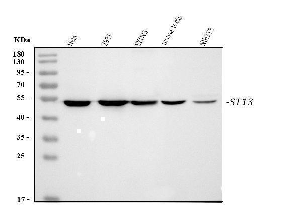
Figure 1. Western blot analysis of ST13 using anti-ST13 antibody (PA1935). Electrophoresis was performed on a 5-20% SDS-PAGE gel at 70V (Stacking gel) / 90V (Resolving gel) for 2-3 hours. The sample well of each lane was loaded with 30 ug of sample under reducing conditions. Lane 1: human Hela whole cell lysates, Lane 2: human 293T whole cell lysates, Lane 3: human SKOV3 whole cell lysates, Lane 4: mouse testis tissue lysates, Lane 5: mouse NIH/3T3 whole cell lysates. After electrophoresis, proteins were transferred to a nitrocellulose membrane at 150 mA for 50-90 minutes. Blocked the membrane with 5% non-fat milk/TBS for 1.5 hour at RT. The membrane was incubated with rabbit anti-ST13 antigen affinity purified polyclonal antibody (Catalog # PA1935) at 0.5 microg/mL overnight at 4°C, then washed with TBS-0.1%Tween 3 times with 5 minutes each and probed with a goat anti-rabbit IgG-HRP secondary antibody at a dilution of 1:5000 for 1.5 hour at RT. The signal is developed using an Enhanced Chemiluminescent detection (ECL) kit (Catalog # EK1002) with Tanon 5200 system. A specific band was detected for ST13 at approximately 45-54 kDa. The expected band size for ST13 is at 41 kDa.
Anti-HSC70 Interacting Protein HIP/ST13 Antibody Picoband(r)

PA1935-DYLIGHT594
ApplicationsImmunoFluorescence, Western Blot, ImmunoCytoChemistry
Product group Antibodies
ReactivityHamster, Human
TargetST13
Overview
- SupplierBoster Bio
- Product NameAnti-HSC70 Interacting Protein HIP/ST13 Antibody Picoband(r)
- Delivery Days Customer9
- Antibody SpecificityNo cross reactivity with other proteins.
- Application Supplier NoteTested Species: In-house tested species with positive results. Predicted Species: Species predicted to be fit for the product based on sequence similarities. Other applications have not been tested. Optimal dilutions should be determined by end users.
- ApplicationsImmunoFluorescence, Western Blot, ImmunoCytoChemistry
- CertificationResearch Use Only
- ClonalityPolyclonal
- Concentration500 ug/ml
- ConjugateOther Conjugate
- Gene ID6767
- Target nameST13
- Target descriptionST13 Hsp70 interacting protein
- Target synonymsAAG2; aging-associated protein 2; FAM10A1; FAM10A4; heat shock 70kD protein binding protein; HIP; HOP; hsc70-interacting protein; Hsp70-interacting protein; HSPABP; HSPABP1; P48; PRO0786; progesterone receptor-associated p48 protein; putative tumor suppressor ST13; renal carcinoma antigen NY-REN-33; SNC6; suppression of tumorigenicity 13 (colon carcinoma) (Hsp70 interacting protein); suppression of tumorigenicity 13 protein; testis secretory sperm-binding protein Li 233m
- HostRabbit
- IsotypeIgG
- Protein IDP50502
- Protein NameHsc70-interacting protein
- Scientific DescriptionBoster Bio Anti-HSC70 Interacting Protein HIP/ST13 Antibody catalog # PA1935. Tested in IF, ICC, WB applications. This antibody reacts with Human. The brand Picoband indicates this is a premium antibody that guarantees superior quality, high affinity, and strong signals with minimal background in Western blot applications. Only our best-performing antibodies are designated as Picoband, ensuring unmatched performance.
- ReactivityHamster, Human
- Storage Instruction-20°C,2°C to 8°C
- UNSPSC12352203