

Figure 1. Western blot analysis of HSD11B1 using anti-HSD11B1 antibody (PA1372-1). Electrophoresis was performed on a 5-20% SDS-PAGE gel at 70V (Stacking gel) / 90V (Resolving gel) for 2-3 hours. The sample well of each lane was loaded with 30 ug of sample under reducing conditions. Lane 1: human hepatocellular carcinoma tumor tissue (HCCT) lysates, Lane 2: human hepatocellular carcinoma paracancerous tissue (HCCP) lysates, Lane 3: monkey liver tissue lysates, Lane 4: rat liver tissue lysates, Lane 5: mouse liver tissue lysates. After electrophoresis, proteins were transferred to a nitrocellulose membrane at 150 mA for 50-90 minutes. Blocked the membrane with 5% non-fat milk/TBS for 1.5 hour at RT. The membrane was incubated with rabbit anti-HSD11B1 antigen affinity purified polyclonal antibody (Catalog # PA1372-1) at 0.5 microg/mL overnight at 4°C, then washed with TBS-0.1%Tween 3 times with 5 minutes each and probed with a goat anti-rabbit IgG-HRP secondary antibody at a dilution of 1:5000 for 1.5 hour at RT. The signal is developed using an Enhanced Chemiluminescent detection (ECL) kit (Catalog # EK1002) with Tanon 5200 system. A specific band was detected for HSD11B1 at approximately 36 kDa. The expected band size for HSD11B1 is at 32 kDa.
. HSD11B1 was detected in a paraffin-embedded section of human liver cancer tissue. Heat mediated antigen retrieval was performed in EDTA buffer (pH 8.0, epitope retrieval solution). The tissue section was blocked with 10% goat serum. The tissue section was then incubated with 2 microg/ml rabbit anti-HSD11B1 Antibody (PA1372-1) overnight at 4°C. Peroxidase Conjugated Goat Anti-rabbit IgG was used as secondary antibody and incubated for 30 minutes at 37°C. The tissue section was developed using HRP Conjugated Rabbit IgG Super Vision Assay Kit (Catalog # SV0002) with DAB as the chromogen.")
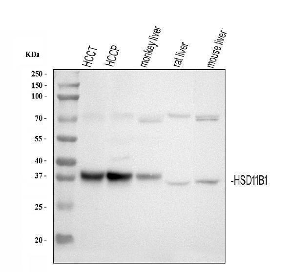
Figure 1. Western blot analysis of HSD11B1 using anti-HSD11B1 antibody (PA1372-1). Electrophoresis was performed on a 5-20% SDS-PAGE gel at 70V (Stacking gel) / 90V (Resolving gel) for 2-3 hours. The sample well of each lane was loaded with 30 ug of sample under reducing conditions. Lane 1: human hepatocellular carcinoma tumor tissue (HCCT) lysates, Lane 2: human hepatocellular carcinoma paracancerous tissue (HCCP) lysates, Lane 3: monkey liver tissue lysates, Lane 4: rat liver tissue lysates, Lane 5: mouse liver tissue lysates. After electrophoresis, proteins were transferred to a nitrocellulose membrane at 150 mA for 50-90 minutes. Blocked the membrane with 5% non-fat milk/TBS for 1.5 hour at RT. The membrane was incubated with rabbit anti-HSD11B1 antigen affinity purified polyclonal antibody (Catalog # PA1372-1) at 0.5 microg/mL overnight at 4°C, then washed with TBS-0.1%Tween 3 times with 5 minutes each and probed with a goat anti-rabbit IgG-HRP secondary antibody at a dilution of 1:5000 for 1.5 hour at RT. The signal is developed using an Enhanced Chemiluminescent detection (ECL) kit (Catalog # EK1002) with Tanon 5200 system. A specific band was detected for HSD11B1 at approximately 36 kDa. The expected band size for HSD11B1 is at 32 kDa.
Anti-HSD11B1 Antibody Picoband(r)

PA1372-1
ApplicationsWestern Blot, ImmunoHistoChemistry
Product group Antibodies
ReactivityBovine, Human, Monkey, Mouse, Rat
TargetHSD11B1
Overview
- SupplierBoster Bio
- Product NameAnti-HSD11B1 Antibody
- Delivery Days Customer9
- Antibody SpecificityNo cross reactivity with other proteins.
- ApplicationsWestern Blot, ImmunoHistoChemistry
- Applications SupplierIHP, WB, IHC
- CertificationResearch Use Only
- ClonalityPolyclonal
- Concentration500 ug/ml
- FormulationLyophilized
- Gene ID3290
- Target nameHSD11B1
- Target descriptionhydroxysteroid 11-beta dehydrogenase 1
- Target synonyms11-beta-HSD1; 11-DH; corticosteroid 11-beta-dehydrogenase isozyme 1; CORTRD2; HDL; HSD11; HSD11B; HSD11L; SDR26C1; short chain dehydrogenase/reductase family 26C member 1
- HostRabbit
- IsotypeIgG
- Protein IDP28845
- Protein NameCorticosteroid 11-beta-dehydrogenase isozyme 1
- Scientific DescriptionBoster Bio Anti-HSD11B1 Antibody catalog # PA1372-1. Tested in IHC, WB applications. This antibody reacts with Human, Monkey, Mouse, Rat. The brand Picoband indicates this is a premium antibody that guarantees superior quality, high affinity, and strong signals with minimal background in Western blot applications. Only our best-performing antibodies are designated as Picoband, ensuring unmatched performance.
- ReactivityBovine, Human, Monkey, Mouse, Rat
- Reactivity SupplierHuman, Mouse, Rat, Bovine
- Storage Instruction-20°C,2°C to 8°C
- UNSPSC12352203