


Figure 1. Western blot analysis of HAS1 using anti-HAS1 antibody (A04784-1). Electrophoresis was performed on a 5-20% SDS-PAGE gel at 70V (Stacking gel) / 90V (Resolving gel) for 2-3 hours. The sample well of each lane was loaded with 50ug of sample under reducing conditions. Lane 1: human SHG-44 whole cell lysates, Lane 2: human THP-1 whole cell lysates, Lane 3: rat brain tissue lysates, Lane 4: rat smooth muscle tissue lysates, Lane 5: rat ovary tissue lysates, Lane 6: mouse brain tissue lysates, Lane 7: mouse smooth muscle tissue lysates, Lane 8: mouse ovary tissue lysates, Lane 9: mouse small intestine tissue lysates, Lane 10: mouse Neuro-2a whole cell lysates. After Electrophoresis, proteins were transferred to a Nitrocellulose membrane at 150mA for 50-90 minutes. Blocked the membrane with 5% Non-fat Milk/ TBS for 1.5 hour at RT. The membrane was incubated with rabbit anti-HAS1 antigen affinity purified polyclonal antibody (Catalog # A04784-1) at 0.5 microg/mL overnight at 4°C, then washed with TBS-0.1%Tween 3 times with 5 minutes each and probed with a goat anti-rabbit IgG-HRP secondary antibody at a dilution of 1:10000 for 1.5 hour at RT. The signal is developed using an Enhanced Chemiluminescent detection (ECL) kit (Catalog # EK1002) with Tanon 5200 system. A specific band was detected for HAS1 at approximately 70KD. The expected band size for HAS1 is at 65KD.
. HAS1 was detected in paraffin-embedded section of human lung cancer tissue. Heat mediated antigen retrieval was performed in citrate buffer (pH6, epitope retrieval solution) for 20 mins. The tissue section was blocked with 10% goat serum. The tissue section was then incubated with 1ugmicrog/ml rabbit anti-HAS1 Antibody (A04784-1) overnight at 4°C. Biotinylated goat anti-rabbit IgG was used as secondary antibody and incubated for 30 minutes at 37°C. The tissue section was developed using Strepavidin-Biotin-Complex (SABC)(Catalog # SA1022) with DAB as the chromogen.")
. HAS1 was detected in paraffin-embedded section of human mammary cancer tissue. Heat mediated antigen retrieval was performed in citrate buffer (pH6, epitope retrieval solution) for 20 mins. The tissue section was blocked with 10% goat serum. The tissue section was then incubated with 1ugmicrog/ml rabbit anti-HAS1 Antibody (A04784-1) overnight at 4°C. Biotinylated goat anti-rabbit IgG was used as secondary antibody and incubated for 30 minutes at 37°C. The tissue section was developed using Strepavidin-Biotin-Complex (SABC)(Catalog # SA1022) with DAB as the chromogen.")
. HAS1 was detected in paraffin-embedded section of human mammary cancer tissue. Heat mediated antigen retrieval was performed in citrate buffer (pH6, epitope retrieval solution) for 20 mins. The tissue section was blocked with 10% goat serum. The tissue section was then incubated with 1ugmicrog/ml rabbit anti-HAS1 Antibody (A04784-1) overnight at 4°C. Biotinylated goat anti-rabbit IgG was used as secondary antibody and incubated for 30 minutes at 37°C. The tissue section was developed using Strepavidin-Biotin-Complex (SABC)(Catalog # SA1022) with DAB as the chromogen.")
. HAS1 was detected in paraffin-embedded section of mouse spleen tissue. Heat mediated antigen retrieval was performed in citrate buffer (pH6, epitope retrieval solution) for 20 mins. The tissue section was blocked with 10% goat serum. The tissue section was then incubated with 1ugmicrog/ml rabbit anti-HAS1 Antibody (A04784-1) overnight at 4°C. Biotinylated goat anti-rabbit IgG was used as secondary antibody and incubated for 30 minutes at 37°C. The tissue section was developed using Strepavidin-Biotin-Complex (SABC)(Catalog # SA1022) with DAB as the chromogen.")
. HAS1 was detected in paraffin-embedded section of rat small intestine tissue. Heat mediated antigen retrieval was performed in citrate buffer (pH6, epitope retrieval solution) for 20 mins. The tissue section was blocked with 10% goat serum. The tissue section was then incubated with 1ugmicrog/ml rabbit anti-HAS1 Antibody (A04784-1) overnight at 4°C. Biotinylated goat anti-rabbit IgG was used as secondary antibody and incubated for 30 minutes at 37°C. The tissue section was developed using Strepavidin-Biotin-Complex (SABC)(Catalog # SA1022) with DAB as the chromogen.")
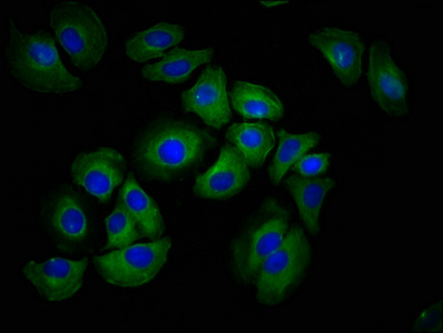
. HAS1 was detected in immunocytochemical section of U20S cell. Enzyme antigen retrieval was performed using IHC enzyme antigen retrieval reagent (AR0022) for 15 mins. The cells were blocked with 10% goat serum. And then incubated with 2microg/mL rabbit anti-HAS1 Antibody (A04784-1) overnight at 4°C. DyLight?488 Conjugated Goat Anti-Rabbit IgG (BA1127) was used as secondary antibody at 1:100 dilution and incubated for 30 minutes at 37°C. The section was counterstained with DAPI. Visualize using a fluorescence microscope and filter sets appropriate for the label used.")
. HAS1 was detected in immunocytochemical section of U20S cell. Enzyme antigen retrieval was performed using IHC enzyme antigen retrieval reagent (AR0022) for 15 mins. The cells were blocked with 10% goat serum. And then incubated with 2microg/mL rabbit anti-HAS1 Antibody (A04784-1) overnight at 4°C. DyLight?488 Conjugated Goat Anti-Rabbit IgG (BA1127) was used as secondary antibody at 1:100 dilution and incubated for 30 minutes at 37°C. The section was counterstained with DAPI. Visualize using a fluorescence microscope and filter sets appropriate for the label used.")
Figure 1. Western blot analysis of HAS1 using anti-HAS1 antibody (A04784-1). Electrophoresis was performed on a 5-20% SDS-PAGE gel at 70V (Stacking gel) / 90V (Resolving gel) for 2-3 hours. The sample well of each lane was loaded with 50ug of sample under reducing conditions. Lane 1: human SHG-44 whole cell lysates, Lane 2: human THP-1 whole cell lysates, Lane 3: rat brain tissue lysates, Lane 4: rat smooth muscle tissue lysates, Lane 5: rat ovary tissue lysates, Lane 6: mouse brain tissue lysates, Lane 7: mouse smooth muscle tissue lysates, Lane 8: mouse ovary tissue lysates, Lane 9: mouse small intestine tissue lysates, Lane 10: mouse Neuro-2a whole cell lysates. After Electrophoresis, proteins were transferred to a Nitrocellulose membrane at 150mA for 50-90 minutes. Blocked the membrane with 5% Non-fat Milk/ TBS for 1.5 hour at RT. The membrane was incubated with rabbit anti-HAS1 antigen affinity purified polyclonal antibody (Catalog # A04784-1) at 0.5 microg/mL overnight at 4°C, then washed with TBS-0.1%Tween 3 times with 5 minutes each and probed with a goat anti-rabbit IgG-HRP secondary antibody at a dilution of 1:10000 for 1.5 hour at RT. The signal is developed using an Enhanced Chemiluminescent detection (ECL) kit (Catalog # EK1002) with Tanon 5200 system. A specific band was detected for HAS1 at approximately 70KD. The expected band size for HAS1 is at 65KD.
Anti-Hyaluronan synthase 1/HAS1 Antibody Picoband(r)
A04784-1
ApplicationsImmunoFluorescence, Western Blot, ImmunoCytoChemistry, ImmunoHistoChemistry
Product group Antibodies
ReactivityHuman, Mouse, Rat
TargetHAS1
Overview
- SupplierBoster Bio
- Product NameAnti-Hyaluronan synthase 1/HAS1 Antibody Picoband(r)
- Delivery Days Customer9
- ApplicationsImmunoFluorescence, Western Blot, ImmunoCytoChemistry, ImmunoHistoChemistry
- CertificationResearch Use Only
- ClonalityPolyclonal
- Concentration500 ug/ml
- Gene ID3036
- Target nameHAS1
- Target descriptionhyaluronan synthase 1
- Target synonymsHAS, hyaluronan synthase 1, HA synthase 1, hyaluronate synthase 1, hyaluronic acid synthase 1
- HostRabbit
- IsotypeIgG
- Protein IDQ92839
- Protein NameHyaluronan synthase 1
- Scientific DescriptionBoster Bio Anti-Hyaluronan synthase 1/HAS1 Antibody Picoband® catalog # A04784-1. Tested in IF, IHC, ICC, WB applications. This antibody reacts with Human, Mouse, Rat. The brand Picoband indicates this is a premium antibody that guarantees superior quality, high affinity, and strong signals with minimal background in Western blot applications. Only our best-performing antibodies are designated as Picoband, ensuring unmatched performance.
- ReactivityHuman, Mouse, Rat
- Storage Instruction-20°C,2°C to 8°C
- UNSPSC12352203
Datasheet
MSDS
Related products

Product group Antibodies
Anti-HAS1 AntibodyA11318
ApplicationsWestern Blot
ReactivityHuman, Mouse
- SizePrice
Product group Antibodies

HAS1 / HAS AntibodyLS-C831068
ApplicationsELISA, ImmunoHistoChemistry
ReactivityHuman, Mouse
TargetHAS1
- SizePrice
Product group Antibodies
HAS1 AntibodyCSB-PA010139LA01HU
ApplicationsImmunoFluorescence, ELISA
ReactivityHuman
TargetHAS1
- SizePrice
Product group Antibodies
Anti-Hyaluronan synthase 1/HAS1 Antibody Picoband(r)A04784-1-CARRIER-FREE
ApplicationsImmunoFluorescence, Western Blot, ImmunoCytoChemistry, ImmunoHistoChemistry
ReactivityHuman, Mouse, Rat
TargetHAS1
- SizePrice
Product group Antibodies
References
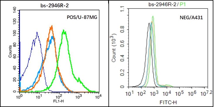
HAS1 Polyclonal AntibodyBS-2946R
ApplicationsFlow Cytometry, ImmunoFluorescence, ELISA, ImmunoCytoChemistry, ImmunoHistoChemistry, ImmunoHistoChemistry Frozen, ImmunoHistoChemistry Paraffin
ReactivityBovine, Human, Mouse, Porcine, Rat, Sheep
TargetHAS1
- SizePrice
Product group Antibodies
HAS1 antibodyGTX04887
ApplicationsWestern Blot
ReactivityHuman, Mouse
TargetHAS1
- SizePrice
Product group Antibodies
Anti-HAS1 Antibody144-61887
ApplicationsWestern Blot
ReactivityHuman, Mouse
TargetHAS1
- SizePrice