

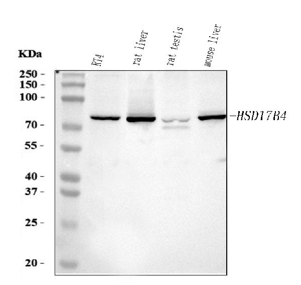
Figure 1. Western blot analysis of HSD17B4 using anti-HSD17B4 antibody (PA1727). Electrophoresis was performed on a 5-20% SDS-PAGE gel at 70V (Stacking gel) / 90V (Resolving gel) for 2-3 hours. The sample well of each lane was loaded with 30 ug of sample under reducing conditions. Lane 1: human RT4 whole cell lysates, Lane 2: rat liver tissue lysates, Lane 3: rat testis tissue lysates, Lane 4: mouse liver tissue lysates. After electrophoresis, proteins were transferred to a nitrocellulose membrane at 150 mA for 50-90 minutes. Blocked the membrane with 5% non-fat milk/TBS for 1.5 hour at RT. The membrane was incubated with rabbit anti-HSD17B4 antigen affinity purified polyclonal antibody (Catalog # PA1727) at 0.5 microg/mL overnight at 4°C, then washed with TBS-0.1%Tween 3 times with 5 minutes each and probed with a goat anti-rabbit IgG-HRP secondary antibody at a dilution of 1:5000 for 1.5 hour at RT. The signal is developed using an Enhanced Chemiluminescent detection (ECL) kit (Catalog # EK1002) with Tanon 5200 system. A specific band was detected for HSD17B4 at approximately 80 kDa. The expected band size for HSD17B4 is at 80 kDa.
. HSD17B4 was detected in a paraffin-embedded section of Human Lung Cancer tissue. Heat mediated antigen retrieval was performed in EDTA buffer (pH 8.0, epitope retrieval solution). The tissue section was blocked with 10% goat serum. The tissue section was then incubated with 1 microg/ml rabbit anti-HSD17B4 Antibody (PA1727) overnight at 4°C. Peroxidase Conjugated Goat Anti-rabbit IgG was used as secondary antibody and incubated for 30 minutes at 37°C. The tissue section was developed using HRP Conjugated Rabbit IgG Super Vision Assay Kit (Catalog # SV0002) with DAB as the chromogen.")
. HSD17B4 was detected in a paraffin-embedded section of Human Intestinal Cancer tissue. Heat mediated antigen retrieval was performed in EDTA buffer (pH 8.0, epitope retrieval solution). The tissue section was blocked with 10% goat serum. The tissue section was then incubated with 1 microg/ml rabbit anti-HSD17B4 Antibody (PA1727) overnight at 4°C. Peroxidase Conjugated Goat Anti-rabbit IgG was used as secondary antibody and incubated for 30 minutes at 37°C. The tissue section was developed using HRP Conjugated Rabbit IgG Super Vision Assay Kit (Catalog # SV0002) with DAB as the chromogen.")
Figure 1. Western blot analysis of HSD17B4 using anti-HSD17B4 antibody (PA1727). Electrophoresis was performed on a 5-20% SDS-PAGE gel at 70V (Stacking gel) / 90V (Resolving gel) for 2-3 hours. The sample well of each lane was loaded with 30 ug of sample under reducing conditions. Lane 1: human RT4 whole cell lysates, Lane 2: rat liver tissue lysates, Lane 3: rat testis tissue lysates, Lane 4: mouse liver tissue lysates. After electrophoresis, proteins were transferred to a nitrocellulose membrane at 150 mA for 50-90 minutes. Blocked the membrane with 5% non-fat milk/TBS for 1.5 hour at RT. The membrane was incubated with rabbit anti-HSD17B4 antigen affinity purified polyclonal antibody (Catalog # PA1727) at 0.5 microg/mL overnight at 4°C, then washed with TBS-0.1%Tween 3 times with 5 minutes each and probed with a goat anti-rabbit IgG-HRP secondary antibody at a dilution of 1:5000 for 1.5 hour at RT. The signal is developed using an Enhanced Chemiluminescent detection (ECL) kit (Catalog # EK1002) with Tanon 5200 system. A specific band was detected for HSD17B4 at approximately 80 kDa. The expected band size for HSD17B4 is at 80 kDa.
Anti-Hydroxysteroid (17-beta) Dehydrogenase 4/HSD17B4 Antibody Picoband(r)

PA1727-DYLIGHT488
ApplicationsWestern Blot, ImmunoHistoChemistry
Product group Antibodies
ReactivityHamster, Human, Mouse, Rat
TargetHSD17B4
Overview
- SupplierBoster Bio
- Product NameAnti-Hydroxysteroid (17-beta) Dehydrogenase 4/HSD17B4 Antibody Picoband(r)
- Delivery Days Customer9
- Antibody SpecificityNo cross reactivity with other proteins.
- Application Supplier NoteTested Species: In-house tested species with positive results. Predicted Species: Species predicted to be fit for the product based on sequence similarities. By Heat: Boiling the paraffin sections in 10mM citrate buffer, pH6.0, for 20mins is required for the staining of formalin/paraffin sections. Other applications have not been tested. Optimal dilutions should be determined by end users.
- ApplicationsWestern Blot, ImmunoHistoChemistry
- CertificationResearch Use Only
- ClonalityPolyclonal
- Concentration500 ug/ml
- ConjugateDyLight 488
- Gene ID3295
- Target nameHSD17B4
- Target descriptionhydroxysteroid 17-beta dehydrogenase 4
- Target synonyms17beta-estradiol dehydrogenase type IV; 17-beta-HSD 4; 17-beta-HSD IV; 17-beta-hydroxysteroid dehydrogenase 4; 3-alpha,7-alpha,12-alpha-trihydroxy-5-beta-cholest-24-enoyl-CoA hydratase; beta-hydroxyacyl dehydrogenase; beta-keto-reductase; D-3-hydroxyacyl-CoA dehydratase; D-bifunctional protein, peroxisomal; DBP; epididymis secretory sperm binding protein; hydroxysteroid dehydrogenase 4; MFE-2; MFP-2; MPF-2; multifunctional protein 2; peroxisomal multifunctional enzyme type 2; peroxisomal multifunctional protein 2; PRLTS1; SDR8C1; short chain dehydrogenase/reductase family 8C member 1
- HostRabbit
- IsotypeIgG
- Protein IDF5HE57
- Protein NamePeroxisomal multifunctional enzyme type 2
- Scientific DescriptionBoster Bio Anti-Hydroxysteroid (17-beta) Dehydrogenase 4/HSD17B4 Antibody catalog # PA1727. Tested in IHC, WB applications. This antibody reacts with Human, Mouse, Rat. The brand Picoband indicates this is a premium antibody that guarantees superior quality, high affinity, and strong signals with minimal background in Western blot applications. Only our best-performing antibodies are designated as Picoband, ensuring unmatched performance.
- ReactivityHamster, Human, Mouse, Rat
- Storage Instruction-20°C,2°C to 8°C
- UNSPSC12352203