

Figure 1. Western blot analysis of IL8 using anti-IL8 antibody (PA1353). Electrophoresis was performed on a 5-20% SDS-PAGE gel at 70V (Stacking gel) / 90V (Resolving gel) for 2-3 hours. Lane 1: recombinant human IL8 protein 10ng. After electrophoresis, proteins were transferred to a nitrocellulose membrane at 150 mA for 50-90 minutes. Blocked the membrane with 5% non-fat milk/TBS for 1.5 hour at RT. The membrane was incubated with rabbit anti-IL8 antigen affinity purified polyclonal antibody (Catalog # PA1353) at 0.5 microg/mL overnight at 4°C, then washed with TBS-0.1%Tween 3 times with 5 minutes each and probed with a goat anti-rabbit IgG-HRP secondary antibody at a dilution of 1:5000 for 1.5 hour at RT. The signal is developed using an Enhanced Chemiluminescent detection (ECL) kit (Catalog # EK1002) with Tanon 5200 system. A specific band was detected for GH1 at approximately 11 kDa.
. IL8 was detected in a paraffin-embedded section of human stomach cancer tissue. Heat mediated antigen retrieval was performed in EDTA buffer (pH 8.0, epitope retrieval solution). The tissue section was blocked with 10% goat serum. The tissue section was then incubated with 2 microg/ml rabbit anti-IL8 Antibody (PA1353) overnight at 4°C. Peroxidase Conjugated Goat Anti-rabbit IgG was used as secondary antibody and incubated for 30 minutes at 37°C. The tissue section was developed using HRP Conjugated Rabbit IgG Super Vision Assay Kit (Catalog # SV0002) with DAB as the chromogen.")
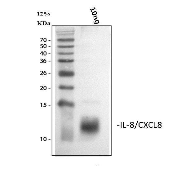
Figure 1. Western blot analysis of IL8 using anti-IL8 antibody (PA1353). Electrophoresis was performed on a 5-20% SDS-PAGE gel at 70V (Stacking gel) / 90V (Resolving gel) for 2-3 hours. Lane 1: recombinant human IL8 protein 10ng. After electrophoresis, proteins were transferred to a nitrocellulose membrane at 150 mA for 50-90 minutes. Blocked the membrane with 5% non-fat milk/TBS for 1.5 hour at RT. The membrane was incubated with rabbit anti-IL8 antigen affinity purified polyclonal antibody (Catalog # PA1353) at 0.5 microg/mL overnight at 4°C, then washed with TBS-0.1%Tween 3 times with 5 minutes each and probed with a goat anti-rabbit IgG-HRP secondary antibody at a dilution of 1:5000 for 1.5 hour at RT. The signal is developed using an Enhanced Chemiluminescent detection (ECL) kit (Catalog # EK1002) with Tanon 5200 system. A specific band was detected for GH1 at approximately 11 kDa.
Anti-IL8/CXCL8 Antibody Picoband(r)

PA1353
ApplicationsWestern Blot, ImmunoHistoChemistry
Product group Antibodies
ReactivityHuman
TargetCXCL8
Overview
- SupplierBoster Bio
- Product NameAnti-IL-8 Antibody
- Delivery Days Customer9
- Antibody SpecificityNo cross reactivity with other proteins.
- Application Supplier NoteTested Species: In-house tested species with positive results. Predicted Species: Species predicted to be fit for the product based on sequence similarities. By Heat: Boiling the paraffin sections in 10mM citrate buffer, pH6.0, for 20mins is required for the staining of formalin/paraffin sections. Other applications have not been tested. Optimal dilutions should be determined by end users.
- ApplicationsWestern Blot, ImmunoHistoChemistry
- Applications SupplierIHP, WB, IHC
- CertificationResearch Use Only
- ClonalityPolyclonal
- Concentration500 ug/ml
- FormulationLyophilized
- Gene ID3576
- Target nameCXCL8
- Target descriptionC-X-C motif chemokine ligand 8
- Target synonymsalveolar macrophage chemotactic factor I; beta endothelial cell-derived neutrophil activating peptide; beta-thromboglobulin-like protein; chemokine (C-X-C motif) ligand 8; emoctakin; GCP1; GCP-1; granulocyte chemotactic protein 1; IL8; interleukin 8; interleukin-8; LECT; LUCT; lung giant cell carcinoma-derived chemotactic protein; lymphocyte derived neutrophil activating peptide; LYNAP; MDNCF; MONAP; monocyte-derived neutrophil chemotactic factor; monocyte-derived neutrophil-activating peptide; NAF; NAP1; NAP-1; neutrophil-activating peptide 1; SCYB8; small inducible cytokine subfamily B, member 8; T-cell chemotactic factor; tumor necrosis factor-induced gene 1
- HostRabbit
- IsotypeIgG
- Protein IDP10145
- Protein NameInterleukin-8
- Scientific DescriptionBoster Bio Anti-IL8/CXCL8 Antibody catalog # PA1353. Tested in IHC, WB applications. This antibody reacts with Human. The brand Picoband indicates this is a premium antibody that guarantees superior quality, high affinity, and strong signals with minimal background in Western blot applications. Only our best-performing antibodies are designated as Picoband, ensuring unmatched performance.
- ReactivityHuman
- Reactivity SupplierHuman
- Storage Instruction-20°C,2°C to 8°C
- UNSPSC12352203