


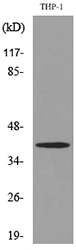
Figure 1. Western blot analysis of IL10 using anti-IL10 antibody (A00021-2). Electrophoresis was performed on a 5-20% SDS-PAGE gel at 70V (Stacking gel) / 90V (Resolving gel) for 2-3 hours. The sample well of each lane was loaded with 50ug of sample under reducing conditions. Lane 1: human U-87MG whole cell lysate, Lane 2: rat testis tissue lysates, Lane 3: mouse testis tissue lysates, Lane 4: mouse SP20 whole cell lysate. After Electrophoresis, proteins were transferred to a Nitrocellulose membrane at 150mA for 50-90 minutes. Blocked the membrane with 5% Non-fat Milk/ TBS for 1.5 hour at RT. The membrane was incubated with rabbit anti-IL10 antigen affinity purified polyclonal antibody (Catalog # A00021-2) at 0.5 microg/mL overnight at 4°C, then washed with TBS-0.1%Tween 3 times with 5 minutes each and probed with a goat anti-rabbit IgG-HRP secondary antibody at a dilution of 1:10000 for 1.5 hour at RT. The signal is developed using an Enhanced Chemiluminescent detection (ECL) kit (Catalog # EK1002) with Tanon 5200 system. A specific band was detected for IL10 at approximately 19KD. The expected band size for IL10 is at 21KD.
Figure 1. Western blot analysis of IL10 using anti-IL10 antibody (A00021-2). Electrophoresis was performed on a 5-20% SDS-PAGE gel at 70V (Stacking gel) / 90V (Resolving gel) for 2-3 hours. The sample well of each lane was loaded with 50ug of sample under reducing conditions. Lane 1: human U-87MG whole cell lysate, Lane 2: rat testis tissue lysates, Lane 3: mouse testis tissue lysates, Lane 4: mouse SP20 whole cell lysate. After Electrophoresis, proteins were transferred to a Nitrocellulose membrane at 150mA for 50-90 minutes. Blocked the membrane with 5% Non-fat Milk/ TBS for 1.5 hour at RT. The membrane was incubated with rabbit anti-IL10 antigen affinity purified polyclonal antibody (Catalog # A00021-2) at 0.5 microg/mL overnight at 4°C, then washed with TBS-0.1%Tween 3 times with 5 minutes each and probed with a goat anti-rabbit IgG-HRP secondary antibody at a dilution of 1:10000 for 1.5 hour at RT. The signal is developed using an Enhanced Chemiluminescent detection (ECL) kit (Catalog # EK1002) with Tanon 5200 system. A specific band was detected for IL10 at approximately 19KD. The expected band size for IL10 is at 21KD.
Anti-IL10 Antibody Picoband(r)
A00021-2
ApplicationsWestern Blot, ELISA
Product group Antibodies
ReactivityHuman, Mouse, Rat
TargetIL10
Overview
- SupplierBoster Bio
- Product NameAnti-IL10 Antibody Picoband(r)
- Delivery Days Customer9
- ApplicationsWestern Blot, ELISA
- CertificationResearch Use Only
- ClonalityPolyclonal
- Concentration500 ug/ml
- Gene ID3586
- Target nameIL10
- Target descriptioninterleukin 10
- Target synonymsCSIF, GVHDS, IL-10, IL10A, TGIF, interleukin-10, T-cell growth inhibitory factor, cytokine synthesis inhibitory factor
- HostRabbit
- IsotypeIgG
- Protein IDP22301
- Protein NameInterleukin-10
- Scientific DescriptionBoster Bio Anti-IL10 Antibody Picoband® catalog # A00021-2. Tested in ELISA, WB applications. This antibody reacts with Human, Mouse, Rat. The brand Picoband indicates this is a premium antibody that guarantees superior quality, high affinity, and strong signals with minimal background in Western blot applications. Only our best-performing antibodies are designated as Picoband, ensuring unmatched performance.
- ReactivityHuman, Mouse, Rat
- Storage Instruction-20°C,2°C to 8°C
- UNSPSC12352203
References
- Jie XL, Tong ZR, Xu XY, et al. Mechanic study based on untargeted metabolomics of Pi-pa-run-fei-tang on pepper combined with ammonia induced chronic cough model mice. J Ethnopharmacol. 2024,326:117905. doi: 10.1016/j.jep.2024.117905Read this paper
- Wang Z, Zhang X, Lv M, et al. Fructus lycii oligosaccharide alleviates acute liver injury via PI3K/Akt/mTOR pathway. Immunol Res. 2024,72(2):271-283. doi: 10.1007/s12026-023-09431-yRead this paper
- Jie XL, Luo ZR, Yu J, et al. Pi-Pa-Run-Fei-Tang alleviates lung injury by modulating IL-6/JAK2/STAT3/IL-17 and PI3K/AKT/NF-κB signaling pathway and balancing Th17 and Treg in murine model of OVA-induced asthma. J Ethnopharmacol. 2023,317:116719. doi: 10.1016/j.jep.2023.116719Read this paper
Datasheet
MSDS
Related products
Product group Antibodies
IL10 AntibodyCSB-PA004870
ApplicationsWestern Blot, ELISA
ReactivityHuman
TargetIL10
- SizePrice
Product group Antibodies
Anti-IL10 Antibody Picoband(r)A00021-2-CARRIER-FREE
ApplicationsWestern Blot, ELISA
ReactivityHuman, Mouse, Rat
TargetIL10
- SizePrice
Product group Antibodies
Anti-IL10 AntibodyA101419
ApplicationsWestern Blot, ELISA
ReactivityHuman
- SizePrice
Product group Antibodies

Anti-IL-10 [JES3-9D7]Ab00139-23.0
ApplicationsWestern Blot, ELISA, ELISpot Assay, ImmunoHistoChemistry, ImmunoHistoChemistry Paraffin, Neutralisation/Blocking
ReactivityHuman, Monkey, Primate
TargetIL10
- SizePrice

Product group Antibodies
ApplicationsELISA, ELISpot Assay
ReactivityHuman
TargetIL10
- SizePrice
Product group Antibodies

IL-10 Antibody (clone JES5-16E3, FITC)LS-C810819
ApplicationsFlow Cytometry
ReactivityMouse
TargetIL10
- SizePrice

Product group Antibodies
Anti-IL10 AntibodyHPA071391
ApplicationsImmunoHistoChemistry
ReactivityHuman
TargetIL10
- SizePrice