

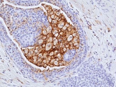
Immunohistochemical staining of formalin-fixed and paraffin-embedded human ductal breast carcinoma tissue sections using anti-LAIR1 antibody (RM498) at 1:100 dilution.
Immunohistochemical staining of formalin-fixed and paraffin-embedded human ductal breast carcinoma tissue sections using anti-LAIR1 antibody (RM498) at 1:100 dilution.
anti-LAIR1 (human), Rabbit Monoclonal (RM498)
REV-31-1390-00
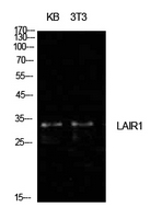
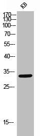
ApplicationsWestern Blot, ImmunoHistoChemistry
Product group Antibodies
ReactivityHuman
TargetLAIR1
Overview
- SupplierRevMAb Biosciences
- Product Nameanti-LAIR1 (human), Rabbit Monoclonal (RM498)
- Delivery Days Customer10
- ApplicationsWestern Blot, ImmunoHistoChemistry
- CertificationResearch Use Only
- ClonalityMonoclonal
- Clone IDRM498
- Gene ID3903
- Target nameLAIR1
- Target descriptionleukocyte associated immunoglobulin like receptor 1
- Target synonymsCD305, LAIR-1, leukocyte-associated immunoglobulin-like receptor 1, immunoglobulin heavy chain variable region, leukocyte-associated Ig-like receptor 1
- HostRabbit
- IsotypeIgG
- Protein IDQ6GTX8
- Protein NameLeukocyte-associated immunoglobulin-like receptor 1
- Scientific DescriptionLAIR1 functions as an inhibitory receptor that plays a constitutive negative regulatory role on the cytolytic function of natural killer (NK) cells, B-cells and T-cells. Activation by Tyr phosphorylation results in the recruitment and activation of the phosphatases PTPN6 and PTPN11. It also reduces the increase of intracellular calcium evoked by B-cell receptor ligation. May also play its inhibitory role independently of SH2-containing phosphatases. Modulates cytokine production in CD4+ T-cells, down-regulating IL2 and IFNG production while inducing secretion of transforming growth factor beta. Down-regulates IgG and IgE production in B-cells as well as IL8, IL10 and TNF secretion. Inhibits proliferation and induces apoptosis in myeloid leukemia cell lines as well as prevents nuclear translocation of NF-kappa-B p65 subunit/RELA and phosphorylation of I-kappa-B alpha/CHUK in these cells. Inhibits the differentiation of peripheral blood precursors towards dendritic cells. LAIR1 plays a role in immune response homeostasis, and extracellular matrix remodeling and it is overexpressed in many high-grade cancers. - Recombinant Antibody. This antibody reacts to human LAIR1. Isotype: Rabbit IgG. Immunogen: Recombinant extracellular domain of human LAIR1. Application: IHC, WB. Liquid. 50% Glycerol/PBS with 1% BSA and 0.09% sodium azide. LAIR1 functions as an inhibitory receptor that plays a constitutive negative regulatory role on the cytolytic function of natural killer (NK) cells, B-cells and T-cells. Activation by Tyr phosphorylation results in the recruitment and activation of the phosphatases PTPN6 and PTPN11. It also reduces the increase of intracellular calcium evoked by B-cell receptor ligation. May also play its inhibitory role independently of SH2-containing phosphatases. Modulates cytokine production in CD4+ T-cells, down-regulating IL2 and IFNG production while inducing secretion of transforming growth factor beta. Down-regulates IgG and IgE production in B-cells as well as IL8, IL10 and TNF secretion. Inhibits proliferation and induces apoptosis in myeloid leukemia cell lines as well as prevents nuclear translocation of NF-kappa-B p65 subunit/RELA and phosphorylation of I-kappa-B alpha/CHUK in these cells. Inhibits the differentiation of peripheral blood precursors towards dendritic cells. LAIR1 plays a role in immune response homeostasis, and extracellular matrix remodeling and it is overexpressed in many high-grade cancers.
- ReactivityHuman
- Storage Instruction-20°C,2°C to 8°C
- UNSPSC41116161
Related products
Product group Antibodies
Anti-LAIR1 AntibodyA100999
ApplicationsWestern Blot, ELISA
ReactivityHuman
- SizePrice

Product group Antibodies
Anti-LAIR1 Antibody Picoband(r)A03470-2-CARRIER-FREE
ApplicationsFlow Cytometry, Western Blot, ELISA
ReactivityHuman
TargetLAIR1
- SizePrice
Product group Antibodies
Anti-LAIR1 Antibody130-10469
ApplicationsWestern Blot, ELISA
TargetLAIR1
- SizePrice
Product group Antibodies

ApplicationsWestern Blot, ELISA
ReactivityHuman
TargetLAIR1
- SizePrice
Product group Antibodies
LAIR1 AntibodyCSB-PA006294
ApplicationsWestern Blot, ELISA
ReactivityHuman
TargetLAIR1
- SizePrice
Product group Antibodies

Goat anti-LAIR1EB09844
ApplicationsWestern Blot, ELISA, ImmunoHistoChemistry
ReactivityHuman
TargetLAIR1
- SizePrice
Product group Antibodies
ApplicationsImmunoPrecipitation, Western Blot, ImmunoCytoChemistry, ImmunoHistoChemistry
TargetLAIR1
- SizePrice
Product group Antibodies
LAIR1 antibody [NKTA255]GTX00485
ApplicationsFlow Cytometry, ImmunoPrecipitation, Western Blot
ReactivityHuman
TargetLAIR1
- SizePrice

Product group Antibodies
Anti-LAIR1 AntibodyHPA011155
ApplicationsWestern Blot, ImmunoCytoChemistry, ImmunoHistoChemistry
ReactivityHuman
TargetLAIR1
- SizePrice
Product group Antibodies
Anti-LAIR1 AntibodyCAB10120
ApplicationsWestern Blot, ELISA
ReactivityHuman
TargetLAIR1
- SizePrice