


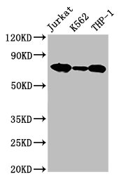
Figure 1. Western blot analysis of LCP1 using anti-LCP1 antibody (A03361). Electrophoresis was performed on a 5-20% SDS-PAGE gel at 70V (Stacking gel) / 90V (Resolving gel) for 2-3 hours. The sample well of each lane was loaded with 30 ug of sample under reducing conditions. Lane 1: human HL-60 whole cell lysates, Lane 2: human THP-1 whole cell lysates, Lane 3: human Hela whole cell lysates, Lane 4: rat spleen tissue lysates, Lane 5: mouse spleen tissue lysates, Lane 6: mouse RAW264.7 whole cell lysates. After electrophoresis, proteins were transferred to a nitrocellulose membrane at 150 mA for 50-90 minutes. Blocked the membrane with 5% non-fat milk/TBS for 1.5 hour at RT. The membrane was incubated with rabbit anti-LCP1 antigen affinity purified polyclonal antibody (Catalog # A03361) at 0.5 microg/mL overnight at 4°C, then washed with TBS-0.1%Tween 3 times with 5 minutes each and probed with a goat anti-rabbit IgG-HRP secondary antibody at a dilution of 1:5000 for 1.5 hour at RT. The signal is developed using an Enhanced Chemiluminescent detection (ECL) kit (Catalog # EK1002) with Tanon 5200 system. A specific band was detected for LCP1 at approximately 70 kDa. The expected band size for LCP1 is at 70 kDa.
. LCP1 was detected in paraffin-embedded section of human B lymphocytic tumor tissue. Heat mediated antigen retrieval was performed in citrate buffer (pH6, epitope retrieval solution) for 20 mins. The tissue section was blocked with 10% goat serum. The tissue section was then incubated with 1microg/ml rabbit anti-LCP1 Antibody (A03361) overnight at 4°C. Biotinylated goat anti-rabbit IgG was used as secondary antibody and incubated for 30 minutes at 37°C. The tissue section was developed using Strepavidin-Biotin-Complex (SABC)(Catalog # SA1022) with DAB as the chromogen.")
. LCP1 was detected in paraffin-embedded section of human tonsil tissue. Heat mediated antigen retrieval was performed in citrate buffer (pH6, epitope retrieval solution) for 20 mins. The tissue section was blocked with 10% goat serum. The tissue section was then incubated with 1microg/ml rabbit anti-LCP1 Antibody (A03361) overnight at 4°C. Biotinylated goat anti-rabbit IgG was used as secondary antibody and incubated for 30 minutes at 37°C. The tissue section was developed using Strepavidin-Biotin-Complex (SABC)(Catalog # SA1022) with DAB as the chromogen.")
. LCP1 was detected in paraffin-embedded section of human tonsil tissue. Heat mediated antigen retrieval was performed in citrate buffer (pH6, epitope retrieval solution) for 20 mins. The tissue section was blocked with 10% goat serum. The tissue section was then incubated with 1microg/ml rabbit anti-LCP1 Antibody (A03361) overnight at 4°C. Biotinylated goat anti-rabbit IgG was used as secondary antibody and incubated for 30 minutes at 37°C. The tissue section was developed using Strepavidin-Biotin-Complex (SABC)(Catalog # SA1022) with DAB as the chromogen.")
. Overlay histogram showing U20S cells stained with A03361 (Blue line). To facilitate intracellular staining, cells were fixed with 4% paraformaldehyde and permeabilized with permeabilization buffer. The cells were blocked with 10% normal goat serum. And then incubated with rabbit anti-LCP1 Antibody (A03361,1microg/1x106 cells) for 30 min at 20°C. DyLight®488 conjugated goat anti-rabbit IgG (BA1127, 5-10microg/1x106 cells) was used as secondary antibody for 30 minutes at 20°C. Isotype control antibody (Green line) was rabbit IgG (1microg/1x106) used under the same conditions. Unlabelled sample (Red line) was also used as a control.")
. LCP1 was detected in immunocytochemical section of A431 cells. Enzyme antigen retrieval was performed using IHC enzyme antigen retrieval reagent (AR0022) for 15 mins. The cells were blocked with 10% goat serum. And then incubated with 2microg/mL rabbit anti-LCP1 Antibody (A03361) overnight at 4°C. DyLight®488 Conjugated Goat Anti-Rabbit IgG (BA1127) was used as secondary antibody at 1:100 dilution and incubated for 30 minutes at 37°C. The section was counterstained with DAPI. Visualize using a fluorescence microscope and filter sets appropriate for the label used.")
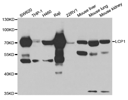
Figure 1. Western blot analysis of LCP1 using anti-LCP1 antibody (A03361). Electrophoresis was performed on a 5-20% SDS-PAGE gel at 70V (Stacking gel) / 90V (Resolving gel) for 2-3 hours. The sample well of each lane was loaded with 30 ug of sample under reducing conditions. Lane 1: human HL-60 whole cell lysates, Lane 2: human THP-1 whole cell lysates, Lane 3: human Hela whole cell lysates, Lane 4: rat spleen tissue lysates, Lane 5: mouse spleen tissue lysates, Lane 6: mouse RAW264.7 whole cell lysates. After electrophoresis, proteins were transferred to a nitrocellulose membrane at 150 mA for 50-90 minutes. Blocked the membrane with 5% non-fat milk/TBS for 1.5 hour at RT. The membrane was incubated with rabbit anti-LCP1 antigen affinity purified polyclonal antibody (Catalog # A03361) at 0.5 microg/mL overnight at 4°C, then washed with TBS-0.1%Tween 3 times with 5 minutes each and probed with a goat anti-rabbit IgG-HRP secondary antibody at a dilution of 1:5000 for 1.5 hour at RT. The signal is developed using an Enhanced Chemiluminescent detection (ECL) kit (Catalog # EK1002) with Tanon 5200 system. A specific band was detected for LCP1 at approximately 70 kDa. The expected band size for LCP1 is at 70 kDa.
Anti-Plastin L/LCP1 Antibody Picoband(r)
A03361
ApplicationsFlow Cytometry, ImmunoFluorescence, Western Blot, ELISA, ImmunoCytoChemistry, ImmunoHistoChemistry
Product group Antibodies
ReactivityHuman, Mouse, Rat
TargetLCP1
Overview
- SupplierBoster Bio
- Product NameAnti-Plastin L/LCP1 Antibody Picoband(r)
- Delivery Days Customer9
- ApplicationsFlow Cytometry, ImmunoFluorescence, Western Blot, ELISA, ImmunoCytoChemistry, ImmunoHistoChemistry
- Applications SupplierWB
- CertificationResearch Use Only
- ClonalityPolyclonal
- Concentration500 ug/ml
- Gene ID3936
- Target nameLCP1
- Target descriptionlymphocyte cytosolic protein 1
- Target synonymsCP64, HEL-S-37, L-PLASTIN, LC64P, LPL, PLS2, plastin-2, L-plastin (Lymphocyte cytosolic protein 1) (LCP-1) (LC64P), LCP-1, Lymphocyte cytosolic protein-1 (plasmin), bA139H14.1 (lymphocyte cytosolic protein 1 (L-plastin)), epididymis secretory protein Li 37
- HostRabbit
- IsotypeIgG
- Protein IDP13796
- Protein NamePlastin-2
- Scientific DescriptionBoster Bio Anti-Plastin L/LCP1 Antibody Picoband® catalog # A03361. Tested in ELISA, Flow Cytometry, IF, IHC, ICC, WB applications. This antibody reacts with Human, Mouse, Rat. The brand Picoband indicates this is a premium antibody that guarantees superior quality, high affinity, and strong signals with minimal background in Western blot applications. Only our best-performing antibodies are designated as Picoband, ensuring unmatched performance.
- ReactivityHuman, Mouse, Rat
- Reactivity SupplierHuman
- Storage Instruction-20°C,2°C to 8°C
- UNSPSC12352203
References
- Yu B, Peng XH, Wang LY, et al. Abnormality of intestinal cholesterol absorption in Apc(Min/+) mice with colon cancer cachexia. Int J Clin Exp Pathol. 2019,12(3):759-767.Read this paper
Datasheet
MSDS
Related products
Product group Antibodies
LCP1 AntibodyCSB-PA012824HA01HU
ApplicationsImmunoPrecipitation, Western Blot, ELISA, ImmunoHistoChemistry
ReactivityHuman
TargetLCP1
- SizePrice
Product group Antibodies
Anti-Plastin L/LCP1 Antibody Picoband(r)A03361-CARRIER-FREE
ApplicationsFlow Cytometry, ImmunoFluorescence, Western Blot, ELISA, ImmunoCytoChemistry, ImmunoHistoChemistry
ReactivityHuman, Mouse, Rat
TargetLCP1
- SizePrice
Product group Antibodies

LCP1 / L-Plastin AntibodyLS-C748551
ApplicationsWestern Blot, ImmunoHistoChemistry
ReactivityHuman, Mouse
TargetLCP1
- SizePrice
Product group Antibodies
Anti-LCP1 AntibodyA30821
ApplicationsImmunoFluorescence, Western Blot, ImmunoHistoChemistry
ReactivityHuman, Mouse, Rat
- SizePrice

Product group Antibodies
Anti-LCP1 AntibodyHPA019493
ApplicationsWestern Blot, ImmunoCytoChemistry, ImmunoHistoChemistry
ReactivityHuman, Rat
TargetLCP1
- SizePrice
Product group Antibodies

ApplicationsWestern Blot, ELISA
ReactivityHuman
TargetLCP1
- SizePrice
Product group Antibodies
Lcp1 Polyclonal AntibodyCAC08589
ApplicationsImmunoFluorescence, Western Blot, ELISA, ImmunoHistoChemistry
ReactivityMouse
TargetLCP1
- SizePrice
Product group Antibodies
L-Plastin antibodyGTX105789
ApplicationsImmunoFluorescence, Western Blot, ImmunoCytoChemistry, ImmunoHistoChemistry, ImmunoHistoChemistry Frozen, ImmunoHistoChemistry Paraffin
ReactivityHuman, Mouse
TargetLCP1
- SizePrice