


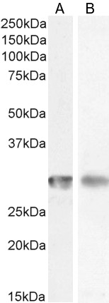
All lanes use the Antibody at 1:2K dilution for 1 hour at room temperature.

. CD4 was detected in a paraffin-embedded section of human appendix tissue. Heat mediated antigen retrieval was performed in EDTA buffer (pH 8.0, epitope retrieval solution). The tissue section was blocked with 10% goat serum. The tissue section was then incubated with 1:50 rabbit anti-CD4 Antibody (M00344) overnight at 4°C. Peroxidase Conjugated Goat Anti-rabbit IgG was used as secondary antibody and incubated for 30 minutes at 37°C. The tissue section was developed using HRP Conjugated Rabbit IgG Super Vision Assay Kit (Catalog # SV0002) with DAB as the chromogen.")
. CD4 was detected in a paraffin-embedded section of human appendix tissue. Heat mediated antigen retrieval was performed in EDTA buffer (pH 8.0, epitope retrieval solution). The tissue section was blocked with 10% goat serum. The tissue section was then incubated with 1:50 rabbit anti-CD4 Antibody (M00344) overnight at 4°C. Peroxidase Conjugated Goat Anti-rabbit IgG was used as secondary antibody and incubated for 30 minutes at 37°C. The tissue section was developed using HRP Conjugated Rabbit IgG Super Vision Assay Kit (Catalog # SV0002) with DAB as the chromogen.")
. CD4 was detected in a paraffin-embedded section of human appendix tissue. Heat mediated antigen retrieval was performed in EDTA buffer (pH 8.0, epitope retrieval solution). The tissue section was blocked with 10% goat serum. The tissue section was then incubated with 1:50 rabbit anti-CD4 Antibody (M00344) overnight at 4°C. Peroxidase Conjugated Goat Anti-rabbit IgG was used as secondary antibody and incubated for 30 minutes at 37°C. The tissue section was developed using HRP Conjugated Rabbit IgG Super Vision Assay Kit (Catalog # SV0002) with DAB as the chromogen.")
. CD4 was detected in a paraffin-embedded section of human appendix tissue. Heat mediated antigen retrieval was performed in EDTA buffer (pH 8.0, epitope retrieval solution). The tissue section was blocked with 10% goat serum. The tissue section was then incubated with 1:50 rabbit anti-CD4 Antibody (M00344) overnight at 4°C. Peroxidase Conjugated Goat Anti-rabbit IgG was used as secondary antibody and incubated for 30 minutes at 37°C. The tissue section was developed using HRP Conjugated Rabbit IgG Super Vision Assay Kit (Catalog # SV0002) with DAB as the chromogen.")
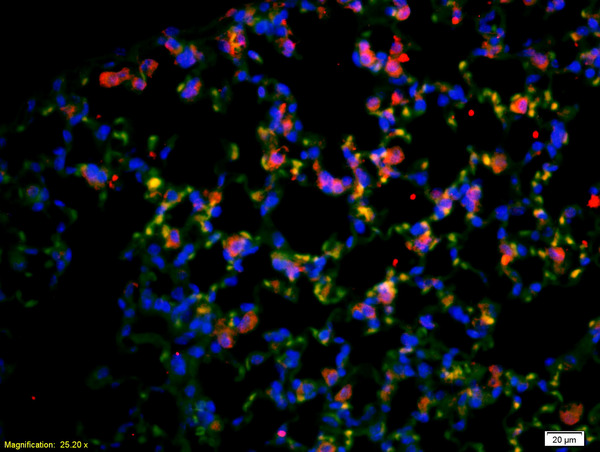
. CD4 was detected in a paraffin-embedded section of human tonsil tissue. Heat mediated antigen retrieval was performed in EDTA buffer (pH 8.0, epitope retrieval solution). The tissue section was blocked with 10% goat serum. The tissue section was then incubated with 1:50 rabbit anti-CD4 Antibody (M00344) overnight at 4°C. Peroxidase Conjugated Goat Anti-rabbit IgG was used as secondary antibody and incubated for 30 minutes at 37°C. The tissue section was developed using HRP Conjugated Rabbit IgG Super Vision Assay Kit (Catalog # SV0002) with DAB as the chromogen.")
. CD4 was detected in a paraffin-embedded section of human tonsil tissue. Heat mediated antigen retrieval was performed in EDTA buffer (pH 8.0, epitope retrieval solution). The tissue section was blocked with 10% goat serum. The tissue section was then incubated with 1:50 rabbit anti-CD4 Antibody (M00344) overnight at 4°C. Peroxidase Conjugated Goat Anti-rabbit IgG was used as secondary antibody and incubated for 30 minutes at 37°C. The tissue section was developed using HRP Conjugated Rabbit IgG Super Vision Assay Kit (Catalog # SV0002) with DAB as the chromogen.")

All lanes use the Antibody at 1:2K dilution for 1 hour at room temperature.
Anti-CD4 Rabbit Monoclonal Antibody
M00344
ApplicationsFlow Cytometry, ImmunoFluorescence, Western Blot, ImmunoCytoChemistry, ImmunoHistoChemistry
Product group Antibodies
ReactivityHuman
TargetCD4
Overview
- SupplierBoster Bio
- Product NameAnti-CD4 Rabbit Monoclonal Antibody
- Delivery Days Customer9
- ApplicationsFlow Cytometry, ImmunoFluorescence, Western Blot, ImmunoCytoChemistry, ImmunoHistoChemistry
- CertificationResearch Use Only
- ClonalityMonoclonal
- Clone IDDOE-3
- Gene ID920
- Target nameCD4
- Target descriptionCD4 molecule
- Target synonymsCD4mut, IMD79, Leu-3, OKT4D, T4, T-cell surface glycoprotein CD4, CD4 antigen (p55), CD4 receptor, T-cell surface antigen T4/Leu-3
- HostRabbit
- IsotypeIgG
- Protein IDP01730
- Protein NameT-cell surface glycoprotein CD4
- Scientific DescriptionBoster Bio Anti-CD4 Rabbit Monoclonal Antibody catalog # M00344. Tested in WB, IHC, ICC/IF, Flow Cytometry applications. This antibody reacts with Human.
- ReactivityHuman
- Storage Instruction-20°C
- UNSPSC12352203
References
- Zhang X, Zhao Q, Cao M, et al. Folate Receptor 4-Expressing T cell Is Associated with Disease-Free Survival in Patients with Esophageal Squamous Cell Carcinoma. Dis Markers. 2022,2022:4351949. doi: 10.1155/2022/4351949Read this paper
- Zhao J, Liu H, Zhang X, et al. Tumor Cells Interleukin-22 Expression Associates with Elevated Tumor-Associated Macrophages Infiltrating and Poor Prognosis in Patients with Breast Cancer. Cancer Biother Radiopharm. 2021,36(2):160-166. doi: 10.1089/cbr.2020.3794Read this paper
- Zhou R, Yang B, Wu X, et al. MicroRNA-421 mediates immunosensitivity in late-stage human liver cancer. Int J Clin Exp Pathol. 2017,10(8):8510-8514.Read this paper
- Zhu HL, Liu YL, Xie XL, et al. Effect of L-arginine on intestinal mucosal immune barrier function in weaned pigs after Escherichia coli LPS challenge. Innate Immun. 2013,19(3):242-52. doi: 10.1177/1753425912456223Read this paper
- Chen X, Wan J, Liu J, et al. Increased IL-17-producing cells correlate with poor survival and lymphangiogenesis in NSCLC patients. Lung Cancer. 2010,69(3):348-54. doi: 10.1016/j.lungcan.2009.11.013Read this paper
Datasheet
MSDS
Related products
Product group Antibodies
Anti-CD4 AntibodyA82715
ApplicationsFlow Cytometry, Western Blot, ELISA, ImmunoHistoChemistry
ReactivityHuman
- SizePrice
Product group Antibodies

Anti-CD4 [YNB46.1.8 (Campath-9H)]Ab00167-23.0
ApplicationsFlow Cytometry, ImmunoHistoChemistry, ImmunoHistoChemistry Frozen
ReactivityHuman
TargetCD4
- SizePrice

Product group Antibodies
ApplicationsFlow Cytometry, ImmunoCytoChemistry
ReactivityHuman
TargetCD4
- SizePrice

Product group Antibodies
Anti-CD4 AntibodyAMAB90754
ApplicationsWestern Blot, ImmunoHistoChemistry
ReactivityHuman
TargetCD4
- SizePrice
Product group Antibodies

CD4 Antibody (clone RPA-T4, FITC)LS-C764187
ApplicationsFlow Cytometry
ReactivityHuman
TargetCD4
- SizePrice
Product group Antibodies
References
CD4 Polyclonal AntibodyBS-0647R
ApplicationsFlow Cytometry, ImmunoFluorescence, Western Blot, ELISA, ImmunoCytoChemistry, ImmunoHistoChemistry, ImmunoHistoChemistry Frozen, ImmunoHistoChemistry Paraffin
ReactivityBovine, Canine, Chicken, Guinea Pig, Human, Mouse, Porcine, Rat, Sheep
TargetCD4
- SizePrice
Product group Antibodies
CD4 Monoclonal AntibodyCSB-MA000228
ApplicationsELISA, ImmunoHistoChemistry
ReactivityHuman, Mouse, Rat
TargetCD4
- SizePrice
Product group Antibodies

Goat anti-CD4EB12597
ApplicationsFlow Cytometry, Western Blot, ELISA, ImmunoHistoChemistry
ReactivityHuman
TargetCD4
- SizePrice
Product group Antibodies
ApplicationsFlow Cytometry
TargetCD4
- SizePrice
![FACS analysis of human peripheral blood lymphocytes using GTX01461-06 CD4 antibody [SK3] (FITC). Solid lone : primary antibody Dashed line : isotype control antibody amount : 0.06 μg (5 μl)](https://www.genetex.com/upload/website/prouct_img/normal/GTX01461-06/GTX01461-06_20200428_FACS68_w_23053121_794.webp)
Product group Antibodies
CD4 antibody [SK3] (FITC)GTX01461-06
ApplicationsFlow Cytometry
ReactivityHuman, Monkey, Primate
TargetCD4
- SizePrice