

Immunohistochemical staining of formalin fixed and paraffin embedded human thymus using Anti-CD28 Rabbit Monoclonal Antibody (Clone RM404) at a 1:200 dilution.
Immunohistochemical staining of formalin fixed and paraffin embedded human thymus using Anti-CD28 Rabbit Monoclonal Antibody (Clone RM404) at a 1:200 dilution.
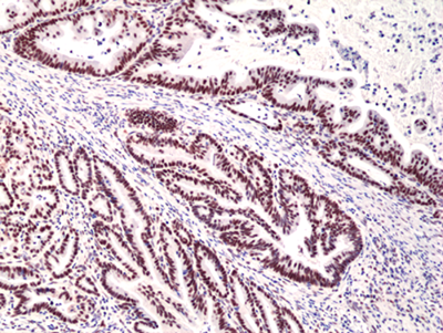
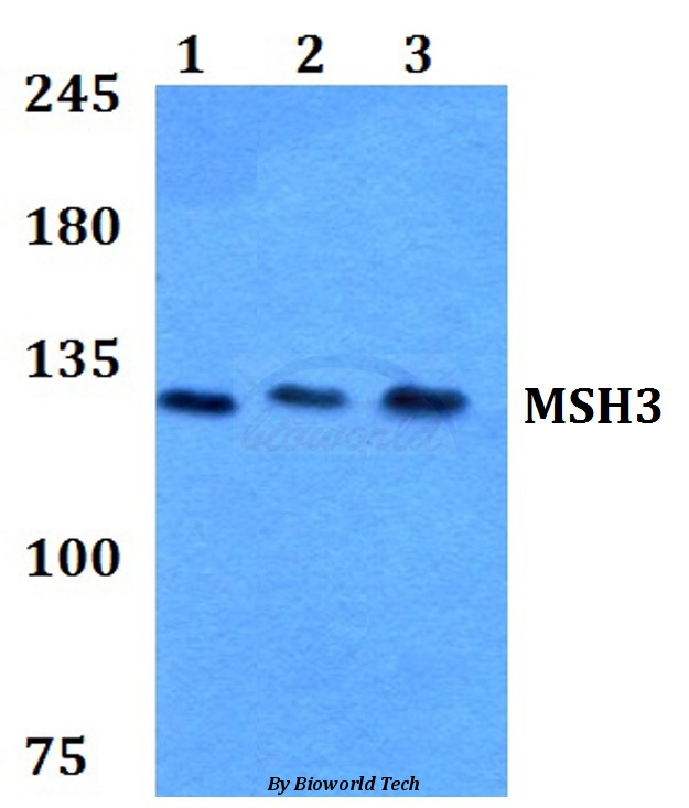
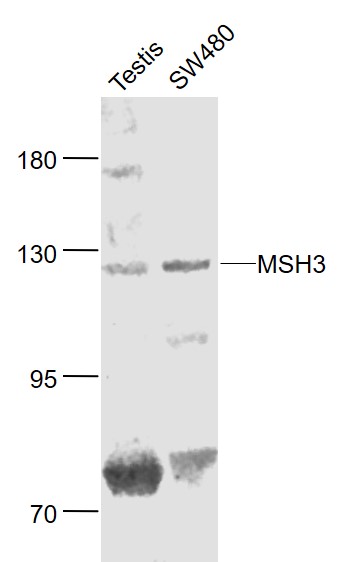
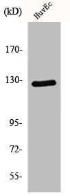
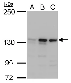
anti-MSH3 (human), Rabbit Monoclonal (RM405)
REV-31-1291-00
ApplicationsWestern Blot, ImmunoHistoChemistry
Product group Antibodies
ReactivityHuman
TargetMSH3
Overview
- SupplierRevMAb Biosciences
- Product Nameanti-MSH3 (human), Rabbit Monoclonal (RM405)
- Delivery Days Customer2
- ApplicationsWestern Blot, ImmunoHistoChemistry
- CertificationResearch Use Only
- ClonalityMonoclonal
- Clone IDRM405
- Gene ID4437
- Target nameMSH3
- Target descriptionmutS homolog 3
- Target synonymsDUP, FAP4, MRP1, DNA mismatch repair protein Msh3, divergent upstream protein, epididymis secretory sperm binding protein, hMSH3, mismatch repair protein 1
- HostRabbit
- IsotypeIgG
- Protein IDP20585
- Protein NameDNA mismatch repair protein Msh3
- Scientific DescriptionRecombinant Antibody. This antibody reacts to human to MSH3. Applications: WB, IHC. Source: Rabbit. Liquid. 50% Glycerol/PBS with 1% BSA and 0.09% sodium azide. The mismatch repair (MMR) proteins are required to maintain genomic integrity in prokaryotes and eukaryotes, by correcting single mismatches and short unpaired regions, such as small insertions and deletions. In eukaryotes, three proteins are involved in mismatch recognition, MSH2, MSH3 and MSH6. The three proteins form two heterodimers MutSalpha (MSH2-MSH6) and MutSbeta (MSH2-MSH3). MutSalpha is thought to be involved primarily in the recognition and repair of base-base mismatches and small insertion/deletion loops. MutSbeta acts preferentially on insertion/deletion loops up to 12 nucleotides in length. The MSH2, MSH3, and PMS2 mismatch repair proteins are also involved in other DNA repair pathways such as single-strand annealing and homologous recombination, anti-recombination, DNA damage signaling, apoptosis, as well as site-specific mutagenesis during immunoglobin somatic hypermutation and class switch recombination. They interact with several other oncogenic targets, including ATR, BRCA1 or p53. Deficiencies in expression of DNA repair genes underlie many forms of cancer. If DNA repair is deficient, DNA damage tends to accumulate. Such excess DNA damage may increase mutations due to error-prone translesion synthesis and error prone repair. Elevated DNA damage may also increase epigenetic alterations due to errors during DNA repair. Such mutations and epigenetic alterations may give rise to cancer. Loss of MSH3 can lead to mismatch repair deficiency and genetic instability which have been identified as particularly common carcinogenic effects in human colorectal cancer. Mutations causing MSH3 knockdown can lead to diminished capacity for cells to repair long insertion/deletion loops causing microsatellite instabilities (MSI) in the genome and allowing an increase in the rates of somatic mutation. - The mismatch repair (MMR) proteins are required to maintain genomic integrity in prokaryotes and eukaryotes, by correcting single mismatches and short unpaired regions, such as small insertions and deletions. In eukaryotes, three proteins are involved in mismatch recognition, MSH2, MSH3 and MSH6. The three proteins form two heterodimers MutSalpha (MSH2-MSH6) and MutSbeta (MSH2-MSH3). MutSalpha is thought to be involved primarily in the recognition and repair of base-base mismatches and small insertion/deletion loops. MutSbeta acts preferentially on insertion/deletion loops up to 12 nucleotides in length. The MSH2, MSH3, and PMS2 mismatch repair proteins are also involved in other DNA repair pathways such as single-strand annealing and homologous recombination, anti-recombination, DNA damage signaling, apoptosis, as well as site-specific mutagenesis during immunoglobin somatic hypermutation and class switch recombination. They interact with several other oncogenic targets, including ATR, BRCA1 or p53. Deficiencies in expression of DNA repair genes underlie many forms of cancer. If DNA repair is deficient, DNA damage tends to accumulate. Such excess DNA damage may increase mutations due to error-prone translesion synthesis and error prone repair. Elevated DNA damage may also increase epigenetic alterations due to errors during DNA repair. Such mutations and epigenetic alterations may give rise to cancer. Loss of MSH3 can lead to mismatch repair deficiency and genetic instability which have been identified as particularly common carcinogenic effects in human colorectal cancer. Mutations causing MSH3 knockdown can lead to diminished capacity for cells to repair long insertion/deletion loops causing microsatellite instabilities (MSI) in the genome and allowing an increase in the rates of somatic mutation.
- ReactivityHuman
- Storage Instruction-20°C,2°C to 8°C
- UNSPSC41116161
Datasheet
Related products
Product group Antibodies
Anti-MSH3 (A80) AntibodyA26260
ApplicationsWestern Blot, ImmunoHistoChemistry
ReactivityHuman, Mouse, Rat
- SizePrice

Product group Antibodies
Anti-MSH3 (A80) AntibodyA02455
ApplicationsWestern Blot, ImmunoHistoChemistry
ReactivityHuman, Mouse, Rat
TargetMSH3
- SizePrice

Product group Antibodies
Anti-MSH3 AntibodyAMAB91910
ApplicationsWestern Blot, ImmunoCytoChemistry, ImmunoHistoChemistry
ReactivityHuman
TargetMSH3
- SizePrice
Product group Antibodies
MSH3 Polyclonal AntibodyBS-4919R
ApplicationsImmunoFluorescence, Western Blot, ImmunoHistoChemistry, ImmunoHistoChemistry Frozen, ImmunoHistoChemistry Paraffin
ReactivityBovine, Equine, Human, Mouse, Porcine, Rabbit, Rat
TargetMSH3
- SizePrice
Product group Antibodies
MSH3 AntibodyCSB-PA003320
ApplicationsWestern Blot, ELISA, ImmunoHistoChemistry
ReactivityHuman
TargetMSH3
- SizePrice
Product group Antibodies
MSH3 antibody [N1N2], N-termGTX103228
ApplicationsWestern Blot
ReactivityHuman
TargetMSH3
- SizePrice
Product group Antibodies

MSH3 AntibodyLS-C334210
ApplicationsWestern Blot, ImmunoHistoChemistry
ReactivityHuman
TargetMSH3
- SizePrice