

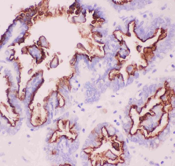
Figure 1. IHC analysis of MUC1 using anti-MUC1 antibody (PB9145). MUC1 was detected in a paraffin-embedded section of human ovary cancer tissue. Heat mediated antigen retrieval was performed in EDTA buffer (pH 8.0, epitope retrieval solution). The tissue section was blocked with 10% goat serum. The tissue section was then incubated with 1 microg/ml rabbit anti-MUC1 Antibody (PB9145) overnight at 4°C. Biotinylated goat anti-rabbit IgG was used as secondary antibody and incubated for 30 minutes at 37°C. The tissue section was developed using Strepavidin-Biotin-Complex (SABC) (Catalog # SA1022) with DAB as the chromogen.
Figure 1. IHC analysis of MUC1 using anti-MUC1 antibody (PB9145). MUC1 was detected in a paraffin-embedded section of human ovary cancer tissue. Heat mediated antigen retrieval was performed in EDTA buffer (pH 8.0, epitope retrieval solution). The tissue section was blocked with 10% goat serum. The tissue section was then incubated with 1 microg/ml rabbit anti-MUC1 Antibody (PB9145) overnight at 4°C. Biotinylated goat anti-rabbit IgG was used as secondary antibody and incubated for 30 minutes at 37°C. The tissue section was developed using Strepavidin-Biotin-Complex (SABC) (Catalog # SA1022) with DAB as the chromogen.
Anti-MUC1 Antibody

PB9145
ApplicationsImmunoHistoChemistry
Product group Antibodies
ReactivityHuman
TargetMUC1
Overview
- SupplierBoster Bio
- Product NameAnti-MUC1 Picoband Antibody
- Delivery Days Customer9
- Antibody SpecificityNo cross reactivity with other proteins.
- Application Supplier NoteTested Species: In-house tested species with positive results. By Heat: Boiling the paraffin sections in 10mM citrate buffer, pH6.0, for 20mins is required for the staining of formalin/paraffin sections. Other applications have not been tested. Optimal dilutions should be determined by end users.
- ApplicationsImmunoHistoChemistry
- Applications SupplierIHP, IHC
- CertificationResearch Use Only
- ClonalityPolyclonal
- Concentration500 ug/ml
- FormulationLyophilized
- Gene ID4582
- Target nameMUC1
- Target descriptionmucin 1, cell surface associated
- Target synonymsADMCKD; ADMCKD1; ADTKD2; breast carcinoma-associated antigen DF3; CA 15-3; cancer antigen 15-3; carcinoma-associated mucin; CD227; EMA; episialin; H23 antigen; H23AG; KL-6; krebs von den Lungen-6; MAM6; MCD; MCKD; MCKD1; MUC-1; MUC-1/SEC; MUC-1/X; MUC1/ZD; mucin 1, transmembrane; mucin-1; peanut-reactive urinary mucin; PEM; PEMT; polymorphic epithelial mucin; PUM; tumor associated epithelial mucin; tumor-associated epithelial membrane antigen
- HostRabbit
- IsotypeIgG
- Protein IDP15941
- Protein NameMucin-1
- Scientific DescriptionBoster Bio Anti-MUC1 Antibody Picoband® catalog # PB9145. Tested in IHC applications. This antibody reacts with Human.
- ReactivityHuman
- Reactivity SupplierHuman
- Storage Instruction-20°C,2°C to 8°C
- UNSPSC12352203