

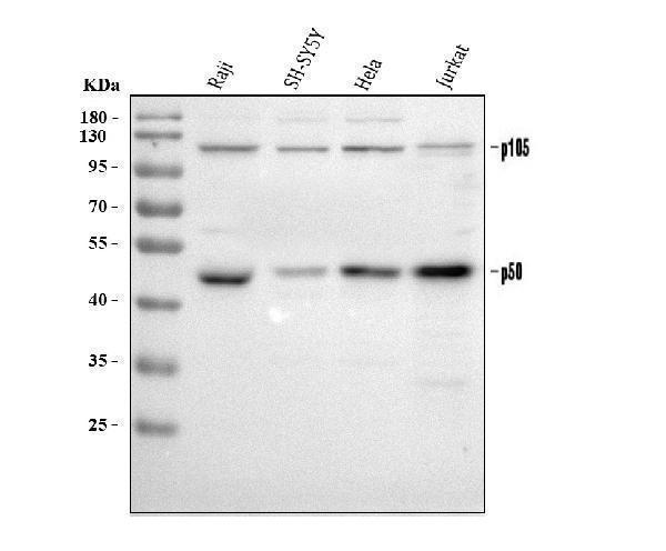
Figure 1. Western blot analysis of NFkB p105/p50/NFKB1 using anti-NFkB p105/p50/NFKB1 antibody (PB9149). Electrophoresis was performed on a 5-20% SDS-PAGE gel at 70V (Stacking gel) / 90V (Resolving gel) for 2-3 hours. The sample well of each lane was loaded with 30 ug of sample under reducing conditions. Lane 1: human Raji whole cell lysates, Lane 2: human SH-SY5Y whole cell lysates, Lane 3: human Hela whole cell lysates, Lane 4: human Jurkat whole cell lysates. After electrophoresis, proteins were transferred to a nitrocellulose membrane at 150 mA for 50-90 minutes. Blocked the membrane with 5% non-fat milk/TBS for 1.5 hour at RT. The membrane was incubated with rabbit anti-NFkB p105/p50/NFKB1 antigen affinity purified polyclonal antibody (Catalog # PB9149) at 0.5 microg/mL overnight at 4°C, then washed with TBS-0.1%Tween 3 times with 5 minutes each and probed with a goat anti-rabbit IgG-HRP secondary antibody at a dilution of 1:5000 for 1.5 hour at RT. The signal is developed using an Enhanced Chemiluminescent detection (ECL) kit (Catalog # EK1002) with Tanon 5200 system. A specific band was detected for NFkB p105/p50/NFKB1 at approximately 50 kDa, 120 kDa. The expected band size for NFkB p105/p50/NFKB1 is at 105 kDa.
. NFkB p105/p50/NFKB1 was detected in an immunocytochemical section of U2OS cells. Enzyme antigen retrieval was performed using IHC enzyme antigen retrieval reagent (AR0022) for 15 mins. The cells were blocked with 10% goat serum. And then incubated with 5 microg/mL rabbit anti-NFkB p105/p50/NFKB1 Antibody (PB9149) overnight at 4°C. Cy3 Conjugated Goat Anti-Rabbit IgG (BA1032) was used as secondary antibody at 1:500 dilution and incubated for 30 minutes at 37°C. The section was counterstained with DAPI. Visualize using a fluorescence microscope and filter sets appropriate for the label used.")
. Overlay histogram showing Hela cells stained with PB9149 (Blue line). To facilitate intracellular staining, cells were fixed with 4% paraformaldehyde and permeabilized with permeabilization buffer. The cells were blocked with 10% normal goat serum. And then incubated with rabbit anti-NFkB p105/p50/NFKB1 Antibody (PB9149, 1 microg/1x106 cells) for 30 min at 20°C. DyLight®488 conjugated goat anti-rabbit IgG (BA1127, 5-10 microg/1x106 cells) was used as secondary antibody for 30 minutes at 20°C. Isotype control antibody (Green line) was rabbit IgG (1 microg/1x106) used under the same conditions. Unlabelled sample without incubation with primary antibody and secondary antibody (Red line) was used as a blank control.")
Figure 1. Western blot analysis of NFkB p105/p50/NFKB1 using anti-NFkB p105/p50/NFKB1 antibody (PB9149). Electrophoresis was performed on a 5-20% SDS-PAGE gel at 70V (Stacking gel) / 90V (Resolving gel) for 2-3 hours. The sample well of each lane was loaded with 30 ug of sample under reducing conditions. Lane 1: human Raji whole cell lysates, Lane 2: human SH-SY5Y whole cell lysates, Lane 3: human Hela whole cell lysates, Lane 4: human Jurkat whole cell lysates. After electrophoresis, proteins were transferred to a nitrocellulose membrane at 150 mA for 50-90 minutes. Blocked the membrane with 5% non-fat milk/TBS for 1.5 hour at RT. The membrane was incubated with rabbit anti-NFkB p105/p50/NFKB1 antigen affinity purified polyclonal antibody (Catalog # PB9149) at 0.5 microg/mL overnight at 4°C, then washed with TBS-0.1%Tween 3 times with 5 minutes each and probed with a goat anti-rabbit IgG-HRP secondary antibody at a dilution of 1:5000 for 1.5 hour at RT. The signal is developed using an Enhanced Chemiluminescent detection (ECL) kit (Catalog # EK1002) with Tanon 5200 system. A specific band was detected for NFkB p105/p50/NFKB1 at approximately 50 kDa, 120 kDa. The expected band size for NFkB p105/p50/NFKB1 is at 105 kDa.
Anti-NFkB/NFKB1 p105/p50 Antibody Picoband(r)

PB9149
ApplicationsFlow Cytometry, ImmunoFluorescence, Western Blot, ImmunoCytoChemistry
Product group Antibodies
ReactivityHuman
TargetNFKB1
Overview
- SupplierBoster Bio
- Product NameAnti-NFkB p105/P50 Picoband Antibody
- Delivery Days Customer9
- Antibody SpecificityNo cross reactivity with other proteins.
- Application Supplier NoteWB: The detection limit for NFkB p105/P50 is approximately 0.25ng/lane under reducing conditions. Tested Species: In-house tested species with positive results. By Heat: Boiling the paraffin sections in 10mM citrate buffer, pH6.0, for 20mins is required for the staining of formalin/paraffin sections. Other applications have not been tested. Optimal dilutions should be determined by end users.
- ApplicationsFlow Cytometry, ImmunoFluorescence, Western Blot, ImmunoCytoChemistry
- Applications SupplierIHP, WB, IHC
- CertificationResearch Use Only
- ClonalityPolyclonal
- Concentration500 ug/ml
- FormulationLyophilized
- Gene ID4790
- Target nameNFKB1
- Target descriptionnuclear factor kappa B subunit 1
- Target synonymsCVID12; DNA-binding factor KBF1; EBP-1; KBF1; NFkappaB; NF-kappaB; NF-kappa-B1; NF-kappabeta; NF-kB; NF-kB1; NFKB-p105; NFKB-p50; nuclear factor kappa-B DNA binding subunit; nuclear factor NF-kappa-B p105 subunit; nuclear factor NF-kappa-B p50 subunit; nuclear factor of kappa light polypeptide gene enhancer in B-cells 1
- HostRabbit
- IsotypeIgG
- Protein IDP19838
- Protein NameNuclear factor NF-kappa-B p105 subunit
- Scientific DescriptionBoster Bio Anti-NFkB/NFKB1 p105/p50 Antibody Picoband® catalog # PB9149. Tested in Flow Cytometry, IF, ICC, WB applications. This antibody reacts with Human. The brand Picoband indicates this is a premium antibody that guarantees superior quality, high affinity, and strong signals with minimal background in Western blot applications. Only our best-performing antibodies are designated as Picoband, ensuring unmatched performance.
- ReactivityHuman
- Reactivity SupplierHuman, Rat
- Storage Instruction-20°C,2°C to 8°C
- UNSPSC12352203
References
- HBOA ameliorates CCl(4)-incuded liver fibrosis through inhibiting TGF-beta1/Smads, NF-kappaB and ERK signaling pathways. Sun X et al., 2019 Jul, Biomed PharmacotherRead more