

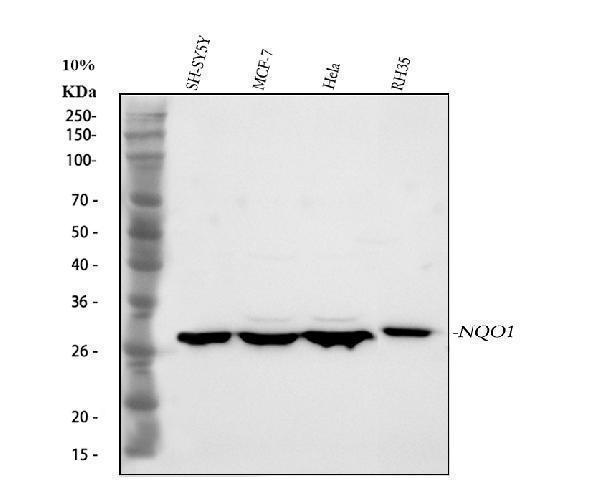
Figure 1. Western blot analysis of NQO1 using anti-NQO1 antibody (PB9497). Electrophoresis was performed on a 5-20% SDS-PAGE gel at 70V (Stacking gel) / 90V (Resolving gel) for 2-3 hours. The sample well of each lane was loaded with 30 ug of sample under reducing conditions. Lane 1: human SH-SY5Y whole cell lysates, Lane 2: human MCF-7 whole cell lysates, Lane 3: human Hela whole cell lysates, Lane 4: rat RH35 whole cell lysates. After electrophoresis, proteins were transferred to a nitrocellulose membrane at 150 mA for 50-90 minutes. Blocked the membrane with 5% non-fat milk/TBS for 1.5 hour at RT. The membrane was incubated with rabbit anti-NQO1 antigen affinity purified polyclonal antibody (Catalog # PB9497) at 0.5 microg/mL overnight at 4°C, then washed with TBS-0.1%Tween 3 times with 5 minutes each and probed with a goat anti-rabbit IgG-HRP secondary antibody at a dilution of 1:5000 for 1.5 hour at RT. The signal is developed using an Enhanced Chemiluminescent detection (ECL) kit (Catalog # EK1002) with Tanon 5200 system. A specific band was detected for NQO1 at approximately 31 kDa. The expected band size for NQO1 is at 31 kDa.
Figure 1. Western blot analysis of NQO1 using anti-NQO1 antibody (PB9497). Electrophoresis was performed on a 5-20% SDS-PAGE gel at 70V (Stacking gel) / 90V (Resolving gel) for 2-3 hours. The sample well of each lane was loaded with 30 ug of sample under reducing conditions. Lane 1: human SH-SY5Y whole cell lysates, Lane 2: human MCF-7 whole cell lysates, Lane 3: human Hela whole cell lysates, Lane 4: rat RH35 whole cell lysates. After electrophoresis, proteins were transferred to a nitrocellulose membrane at 150 mA for 50-90 minutes. Blocked the membrane with 5% non-fat milk/TBS for 1.5 hour at RT. The membrane was incubated with rabbit anti-NQO1 antigen affinity purified polyclonal antibody (Catalog # PB9497) at 0.5 microg/mL overnight at 4°C, then washed with TBS-0.1%Tween 3 times with 5 minutes each and probed with a goat anti-rabbit IgG-HRP secondary antibody at a dilution of 1:5000 for 1.5 hour at RT. The signal is developed using an Enhanced Chemiluminescent detection (ECL) kit (Catalog # EK1002) with Tanon 5200 system. A specific band was detected for NQO1 at approximately 31 kDa. The expected band size for NQO1 is at 31 kDa.
Anti-NQO1 Antibody Picoband(r)

PB9497
ApplicationsWestern Blot
Product group Antibodies
ReactivityHuman, Rat
TargetNQO1
Overview
- SupplierBoster Bio
- Product NameAnti-NQO1 Picoband Antibody
- Delivery Days Customer9
- Antibody SpecificityNo cross reactivity with other proteins.
- Application Supplier NoteTested Species: In-house tested species with positive results. Other applications have not been tested. Optimal dilutions should be determined by end users.
- ApplicationsWestern Blot
- CertificationResearch Use Only
- ClonalityPolyclonal
- Concentration500 ug/ml
- FormulationLyophilized
- Gene ID1728
- Target nameNQO1
- Target descriptionNAD(P)H quinone dehydrogenase 1
- Target synonymsazoreductase; DHQU; DIA4; diaphorase (NADH/NADPH) (cytochrome b-5 reductase); diaphorase-4; dioxin-inducible 1; DTD; DT-diaphorase; menadione reductase; NAD(P)H dehydrogenase [quinone] 1; NAD(P)H dehydrogenase, quinone 1; NAD(P)H:menadione oxidoreductase 1; NAD(P)H:Quinone acceptor oxidoreductase type 1; NAD(P)H:quinone oxidoreductase 1; NAD(P)H:quinone oxireductase; NMOR1; NMORI; phylloquinone reductase; QR1; quinone reductase 1
- HostRabbit
- IsotypeIgG
- Protein IDP15559
- Protein NameNAD(P)H dehydrogenase [quinone] 1
- Scientific DescriptionBoster Bio Anti-NQO1 Antibody Picoband® catalog # PB9497. Tested in WB applications. This antibody reacts with Human, Rat. The brand Picoband indicates this is a premium antibody that guarantees superior quality, high affinity, and strong signals with minimal background in Western blot applications. Only our best-performing antibodies are designated as Picoband, ensuring unmatched performance.
- ReactivityHuman, Rat
- Storage Instruction-20°C,2°C to 8°C
- UNSPSC12352203
References
- Linagliptin protects rat carotid artery from balloon injury and activates the NRF2 antioxidant pathway. Si J et al., 2019 Feb 26, Exp AnimRead more