

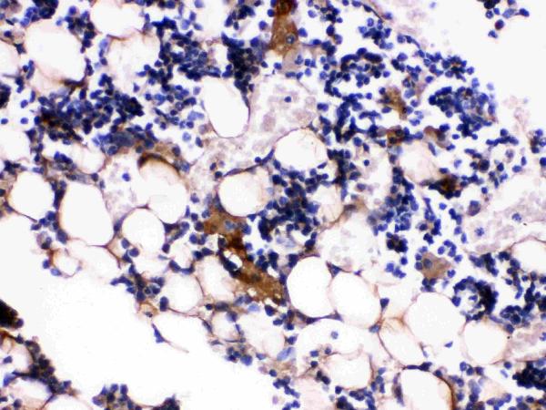
Figure 1. IHC analysis of Osteocalcin using anti-Osteocalcin antibody (PB9919). Osteocalcin was detected in paraffin-embedded section of mouse tibia tissues. Heat mediated antigen retrieval was performed in citrate buffer (pH6, epitope retrieval solution) for 20 mins. The tissue section was blocked with 10% goat serum. The tissue section was then incubated with 1microg/ml rabbit anti-Osteocalcin Antibody (PB9919) overnight at 4°C. Biotinylated goat anti-rabbit IgG was used as secondary antibody and incubated for 30 minutes at 37°C. The tissue section was developed using Strepavidin-Biotin-Complex (SABC)(Catalog # SA1022) with DAB as the chromogen.
Figure 1. IHC analysis of Osteocalcin using anti-Osteocalcin antibody (PB9919). Osteocalcin was detected in paraffin-embedded section of mouse tibia tissues. Heat mediated antigen retrieval was performed in citrate buffer (pH6, epitope retrieval solution) for 20 mins. The tissue section was blocked with 10% goat serum. The tissue section was then incubated with 1microg/ml rabbit anti-Osteocalcin Antibody (PB9919) overnight at 4°C. Biotinylated goat anti-rabbit IgG was used as secondary antibody and incubated for 30 minutes at 37°C. The tissue section was developed using Strepavidin-Biotin-Complex (SABC)(Catalog # SA1022) with DAB as the chromogen.
Anti-Osteocalcin/BGLAP Antibody

PB9919-DYLIGHT550
ApplicationsImmunoHistoChemistry
Product group Antibodies
ReactivityMouse
TargetBglap
Overview
- SupplierBoster Bio
- Product NameAnti-Osteocalcin/BGLAP Antibody
- Delivery Days Customer9
- Antibody SpecificityNo cross reactivity with other proteins.
- Application Supplier NoteTested Species: In-house tested species with positive results. By Heat: Boiling the paraffin sections in 10mM citrate buffer, pH6.0, for 20mins is required for the staining of formalin/paraffin sections. Other applications have not been tested. Optimal dilutions should be determined by end users.
- ApplicationsImmunoHistoChemistry
- CertificationResearch Use Only
- ClonalityPolyclonal
- Concentration500 ug/ml
- ConjugateDyLight 550
- Gene ID12096
- Target nameBglap
- Target descriptionbone gamma carboxyglutamate protein
- Target synonymsBgl; Bglap1; BGP; bone gamma carboxyglutamate protein 1; bone Gla protein; gamma-carboxyglutamic acid-containing protein; mOC; mOC-A; O; OC; OG; OG1; oste; osteocalcin
- HostRabbit
- IsotypeIgG
- Protein IDP86546
- Protein NameOsteocalcin
- Scientific DescriptionBoster Bio Anti-Osteocalcin/BGLAP Antibody Picoband® catalog # PB9919. Tested in IHC applications. This antibody reacts with Mouse.
- ReactivityMouse
- Storage Instruction-20°C,2°C to 8°C
- UNSPSC12352203