


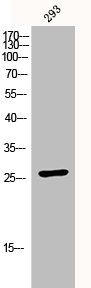
Figure 1. Western blot analysis of CDKN1B using anti-CDKN1B antibody (M00173-1). Electrophoresis was performed on a 5-20% SDS-PAGE gel at 70V (Stacking gel) / 90V (Resolving gel) for 2-3 hours. The sample well of each lane was loaded with 30 ug of sample under reducing conditions. Lane 1: human MCF-7 whole cell lysates, Lane 2: human Jurkat whole cell lysates, Lane 3: human CACO-2 whole cell lysates, Lane 4: human HepG2 whole cell lysates, Lane 5: rat brain tissue lysates, Lane 6: mouse brain tissue lysates. After electrophoresis, proteins were transferred to a nitrocellulose membrane at 150 mA for 50-90 minutes. Blocked the membrane with 5% non-fat milk/TBS for 1.5 hour at RT. The membrane was incubated with rabbit anti-CDKN1B antigen affinity purified monoclonal antibody (Catalog # M00173-1) at 1:500 overnight at 4°C, then washed with TBS-0.1%Tween 3 times with 5 minutes each and probed with a goat anti-rabbit IgG-HRP secondary antibody at a dilution of 1:500 for 1.5 hour at RT. The signal is developed using an Enhanced Chemiluminescent detection (ECL) kit (Catalog # EK1002) with Tanon 5200 system. A specific band was detected for CDKN1B at approximately 25 kDa. The expected band size for CDKN1B is at 22 kDa.
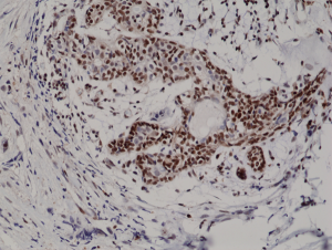
. CDKN1B was detected in a paraffin-embedded section of human breast cancer tissue. Heat mediated antigen retrieval was performed in EDTA buffer (pH 8.0, epitope retrieval solution). The tissue section was blocked with 10% goat serum. The tissue section was then incubated with 1:200 rabbit anti-CDKN1B Antibody (M00173-1) overnight at 4°C. Peroxidase Conjugated Goat Anti-rabbit IgG was used as secondary antibody and incubated for 30 minutes at 37°C. The tissue section was developed using HRP Conjugated Rabbit IgG Super Vision Assay Kit (Catalog # SV0002) with DAB as the chromogen.")
. CDKN1B was detected in a paraffin-embedded section of human lung cancer tissue. Heat mediated antigen retrieval was performed in EDTA buffer (pH 8.0, epitope retrieval solution). The tissue section was blocked with 10% goat serum. The tissue section was then incubated with 1:200 rabbit anti-CDKN1B Antibody (M00173-1) overnight at 4°C. Peroxidase Conjugated Goat Anti-rabbit IgG was used as secondary antibody and incubated for 30 minutes at 37°C. The tissue section was developed using HRP Conjugated Rabbit IgG Super Vision Assay Kit (Catalog # SV0002) with DAB as the chromogen.")
. CDKN1B was detected in a paraffin-embedded section of human urothelial carcinoma with squamous differentiation tissue. Heat mediated antigen retrieval was performed in EDTA buffer (pH 8.0, epitope retrieval solution). The tissue section was blocked with 10% goat serum. The tissue section was then incubated with 1:200 rabbit anti-CDKN1B Antibody (M00173-1) overnight at 4°C. Peroxidase Conjugated Goat Anti-rabbit IgG was used as secondary antibody and incubated for 30 minutes at 37°C. The tissue section was developed using HRP Conjugated Rabbit IgG Super Vision Assay Kit (Catalog # SV0002) with DAB as the chromogen.")
. CDKN1B was detected in a paraffin-embedded section of rat brain tissue. Heat mediated antigen retrieval was performed in EDTA buffer (pH 8.0, epitope retrieval solution). The tissue section was blocked with 10% goat serum. The tissue section was then incubated with 1:200 rabbit anti-CDKN1B Antibody (M00173-1) overnight at 4°C. Peroxidase Conjugated Goat Anti-rabbit IgG was used as secondary antibody and incubated for 30 minutes at 37°C. The tissue section was developed using HRP Conjugated Rabbit IgG Super Vision Assay Kit (Catalog # SV0002) with DAB as the chromogen.")

Figure 1. Western blot analysis of CDKN1B using anti-CDKN1B antibody (M00173-1). Electrophoresis was performed on a 5-20% SDS-PAGE gel at 70V (Stacking gel) / 90V (Resolving gel) for 2-3 hours. The sample well of each lane was loaded with 30 ug of sample under reducing conditions. Lane 1: human MCF-7 whole cell lysates, Lane 2: human Jurkat whole cell lysates, Lane 3: human CACO-2 whole cell lysates, Lane 4: human HepG2 whole cell lysates, Lane 5: rat brain tissue lysates, Lane 6: mouse brain tissue lysates. After electrophoresis, proteins were transferred to a nitrocellulose membrane at 150 mA for 50-90 minutes. Blocked the membrane with 5% non-fat milk/TBS for 1.5 hour at RT. The membrane was incubated with rabbit anti-CDKN1B antigen affinity purified monoclonal antibody (Catalog # M00173-1) at 1:500 overnight at 4°C, then washed with TBS-0.1%Tween 3 times with 5 minutes each and probed with a goat anti-rabbit IgG-HRP secondary antibody at a dilution of 1:500 for 1.5 hour at RT. The signal is developed using an Enhanced Chemiluminescent detection (ECL) kit (Catalog # EK1002) with Tanon 5200 system. A specific band was detected for CDKN1B at approximately 25 kDa. The expected band size for CDKN1B is at 22 kDa.
Anti-p27 KIP 1 CDKN1B Rabbit Monoclonal Antibody
M00173-1
ApplicationsFlow Cytometry, ImmunoFluorescence, ImmunoPrecipitation, Western Blot, ImmunoCytoChemistry, ImmunoHistoChemistry
Product group Antibodies
ReactivityHuman, Mouse, Rat
TargetCDKN1B
Overview
- SupplierBoster Bio
- Product NameAnti-p27 KIP 1 CDKN1B Rabbit Monoclonal Antibody
- Delivery Days Customer9
- ApplicationsFlow Cytometry, ImmunoFluorescence, ImmunoPrecipitation, Western Blot, ImmunoCytoChemistry, ImmunoHistoChemistry
- CertificationResearch Use Only
- ClonalityMonoclonal
- Clone IDCGA-3
- Gene ID1027
- Target nameCDKN1B
- Target descriptioncyclin dependent kinase inhibitor 1B
- Target synonymsCDKN4, KIP1, MEN1B, MEN4, P27KIP1, cyclin-dependent kinase inhibitor 1B, cyclin-dependent kinase inhibitor 1B (p27, Kip1)
- HostRabbit
- IsotypeIgG
- Protein IDP46527
- Protein NameCyclin-dependent kinase inhibitor 1B
- Scientific DescriptionBoster Bio Anti-p27 KIP 1 CDKN1B Rabbit Monoclonal Antibody catalog # M00173-1. Tested in WB, IHC, ICC/IF, IP, Flow Cytometry applications. This antibody reacts with Human, Mouse, Rat.
- ReactivityHuman, Mouse, Rat
- Storage Instruction-20°C
- UNSPSC12352203
References
- Liu Z, Ye Q, Wu L, et al. Metallothionein 1 family profiling identifies MT1X as a tumor suppressor involved in the progression and metastastatic capacity of hepatocellular carcinoma. Mol Carcinog. 2018,57(11):1435-1444. doi: 10.1002/mc.22846Read this paper
- Chen J, Zhuo JY, Yang F, et al. 17-beta-hydroxysteroid dehydrogenase 13 inhibits the progression and recurrence of hepatocellular carcinoma. Hepatobiliary Pancreat Dis Int. 2018,17(3):220-226. doi: 10.1016/j.hbpd.2018.04.006Read this paper
- Fu L, Zhang S. RASSF1A promotes apoptosis and suppresses the proliferation of ovarian cancer cells. Int J Mol Med. 2014,33(5):1153-60. doi: 10.3892/ijmm.2014.1671Read this paper
Datasheet
MSDS
Related products
Product group Antibodies
ReactivityHuman
TargetCDKN1B
- SizePrice
Product group Antibodies
CDKN1B AntibodyCSB-PA003625
ApplicationsWestern Blot, ELISA, ImmunoHistoChemistry
ReactivityHuman, Mouse, Rat
TargetCDKN1B
- SizePrice
Product group Antibodies
ApplicationsWestern Blot, ImmunoHistoChemistry
ReactivityMouse, Rat
TargetCDKN1B
- SizePrice
Product group Antibodies
Anti-p27 Antibody130-10050
ApplicationsELISA
ReactivityHuman
TargetCDKN1B
- SizePrice
Product group Antibodies
Anti-p27 Kip1 AntibodyA95332
ApplicationsWestern Blot, ELISA, ImmunoHistoChemistry
ReactivityHuman, Mouse, Rat
- SizePrice
Product group Antibodies

ApplicationsELISA
ReactivityHuman
TargetCDKN1B
- SizePrice

Product group Antibodies
Anti-CDKN1B AntibodyHPA059086
ApplicationsWestern Blot, ImmunoCytoChemistry
ReactivityHuman
TargetCDKN1B
- SizePrice
Product group Antibodies

ApplicationsImmunoPrecipitation, Western Blot, ELISA
ReactivityBovine, Canine, Human, Mouse, Rat
TargetCDKN1B
- SizePrice
Product group Antibodies
anti-p27Kip1 (human), Rabbit Monoclonal (RM302)REV-31-1187-00
ApplicationsWestern Blot, ImmunoHistoChemistry
ReactivityHuman
TargetCDKN1B
- SizePrice