

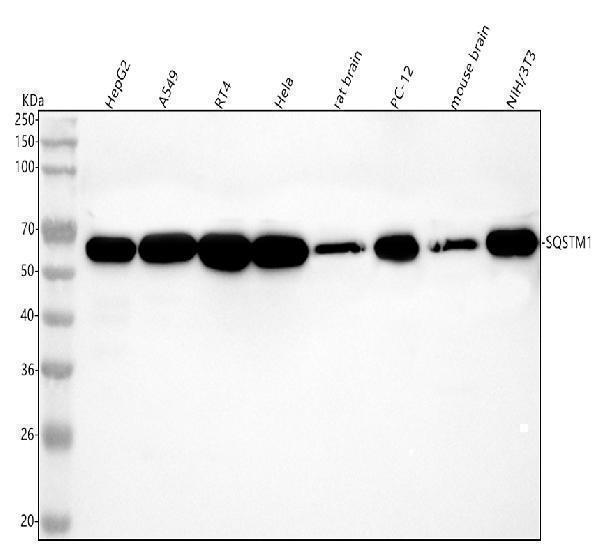
Figure 1. Western blot analysis of p62/SQSTM1 using anti-p62/SQSTM1 antibody (M00300). Electrophoresis was performed on a 5-20% SDS-PAGE gel at 70V (Stacking gel) / 90V (Resolving gel) for 2-3 hours. The sample well of each lane was loaded with 30 ug of sample under reducing conditions. Lane 1: human HepG2 whole cell lysates, Lane 2: human A549 whole cell lysates, Lane 3: human RT4 whole cell lysates, Lane 4: human Hela whole cell lysates, Lane 5: rat brain tissue lysates, Lane 6: rat PC-12 whole cell lysates, Lane 7: mouse brain tissue lysates, Lane 8: mouse NIH/3T3 whole cell lysates. After electrophoresis, proteins were transferred to a nitrocellulose membrane at 150 mA for 50-90 minutes. Blocked the membrane with 5% non-fat milk/TBS for 1.5 hour at RT. The membrane was incubated with rabbit anti-p62/SQSTM1 antigen affinity purified monoclonal antibody (Catalog # M00300) at 1:5000 overnight at 4°C, then washed with TBS-0.1%Tween 3 times with 5 minutes each and probed with a goat anti-rabbit IgG-HRP secondary antibody at a dilution of 1:1000 for 1.5 hour at RT. The signal is developed using an Enhanced Chemiluminescent detection (ECL) kit (Catalog # EK1002) with Tanon 5200 system. A specific band was detected for p62/SQSTM1 at approximately 62 kDa. The expected band size for p62/SQSTM1 is at 48 kDa.
. p62/SQSTM1 was detected in a paraffin-embedded section of human colorectal adenocarcinoma tissue. Heat mediated antigen retrieval was performed in EDTA buffer (pH 8.0, epitope retrieval solution). The tissue section was blocked with 10% goat serum. The tissue section was then incubated with 1:50 rabbit anti-p62/SQSTM1 Antibody (M00300) overnight at 4°C. Peroxidase Conjugated Goat Anti-rabbit IgG was used as secondary antibody and incubated for 30 minutes at 37°C. The tissue section was developed using HRP Conjugated Rabbit IgG Super Vision Assay Kit (Catalog # SV0002) with DAB as the chromogen.")
. p62/SQSTM1 was detected in a paraffin-embedded section of human spleen tissue. Heat mediated antigen retrieval was performed in EDTA buffer (pH 8.0, epitope retrieval solution). The tissue section was blocked with 10% goat serum. The tissue section was then incubated with 1:50 rabbit anti-p62/SQSTM1 Antibody (M00300) overnight at 4°C. Peroxidase Conjugated Goat Anti-rabbit IgG was used as secondary antibody and incubated for 30 minutes at 37°C. The tissue section was developed using HRP Conjugated Rabbit IgG Super Vision Assay Kit (Catalog # SV0002) with DAB as the chromogen.")

. p62/SQSTM1 was detected in a paraffin-embedded section of human cervix squamous cell carcinoma tissue. Heat mediated antigen retrieval was performed in EDTA buffer (pH 8.0, epitope retrieval solution). The tissue section was blocked with 10% goat serum. The tissue section was then incubated with 1:50 rabbit anti-p62/SQSTM1 Antibody (M00300) overnight at 4°C. Peroxidase Conjugated Goat Anti-rabbit IgG was used as secondary antibody and incubated for 30 minutes at 37°C. The tissue section was developed using HRP Conjugated Rabbit IgG Super Vision Assay Kit (Catalog # SV0002) with DAB as the chromogen.")

Figure 1. Western blot analysis of p62/SQSTM1 using anti-p62/SQSTM1 antibody (M00300). Electrophoresis was performed on a 5-20% SDS-PAGE gel at 70V (Stacking gel) / 90V (Resolving gel) for 2-3 hours. The sample well of each lane was loaded with 30 ug of sample under reducing conditions. Lane 1: human HepG2 whole cell lysates, Lane 2: human A549 whole cell lysates, Lane 3: human RT4 whole cell lysates, Lane 4: human Hela whole cell lysates, Lane 5: rat brain tissue lysates, Lane 6: rat PC-12 whole cell lysates, Lane 7: mouse brain tissue lysates, Lane 8: mouse NIH/3T3 whole cell lysates. After electrophoresis, proteins were transferred to a nitrocellulose membrane at 150 mA for 50-90 minutes. Blocked the membrane with 5% non-fat milk/TBS for 1.5 hour at RT. The membrane was incubated with rabbit anti-p62/SQSTM1 antigen affinity purified monoclonal antibody (Catalog # M00300) at 1:5000 overnight at 4°C, then washed with TBS-0.1%Tween 3 times with 5 minutes each and probed with a goat anti-rabbit IgG-HRP secondary antibody at a dilution of 1:1000 for 1.5 hour at RT. The signal is developed using an Enhanced Chemiluminescent detection (ECL) kit (Catalog # EK1002) with Tanon 5200 system. A specific band was detected for p62/SQSTM1 at approximately 62 kDa. The expected band size for p62/SQSTM1 is at 48 kDa.
Anti-p62/SQSTM1 Rabbit Monoclonal Antibody

M00300
ApplicationsFlow Cytometry, ImmunoFluorescence, ImmunoPrecipitation, Western Blot, ImmunoCytoChemistry, ImmunoHistoChemistry
Product group Antibodies
ReactivityHuman, Mouse, Rat
TargetSQSTM1
Overview
- SupplierBoster Bio
- Product NameAnti-p62/SQSTM1 Rabbit Monoclonal Antibody
- Delivery Days Customer9
- ApplicationsFlow Cytometry, ImmunoFluorescence, ImmunoPrecipitation, Western Blot, ImmunoCytoChemistry, ImmunoHistoChemistry
- CertificationResearch Use Only
- ClonalityMonoclonal
- Clone IDEBB-19
- FormulationLiquid
- Gene ID8878
- Target nameSQSTM1
- Target descriptionsequestosome 1
- Target synonymsA170; autophagy receptor p62; DMRV; EBI3-associated protein of 60 kDa; EBI3-associated protein p60; EBIAP; FTDALS3; NADGP; OSIL; oxidative stress induced like; p60; p62; p62B; PDB3; phosphotyrosine independent ligand for the Lck SH2 domain p62; phosphotyrosine-independent ligand for the Lck SH2 domain of 62 kDa; sequestosome-1; ubiquitin-binding protein p62; ZIP3
- HostRabbit
- IsotypeIgG
- Protein IDQ13501
- Protein NameSequestosome-1
- Scientific DescriptionBoster Bio Anti-p62/SQSTM1 Rabbit Monoclonal Antibody catalog # M00300. Tested in WB, IHC, ICC/IF, Flow Cytometry, IP applications. This antibody reacts with Human, Mouse, Rat.
- ReactivityHuman, Mouse, Rat
- Storage Instruction-20°C
- UNSPSC12352203
References
- Integral membrane protein 2A inhibits cell growth in human breast cancer via enhancing autophagy induction. Zhou C et al., 2019 Aug 22, Cell Commun SignalRead more