

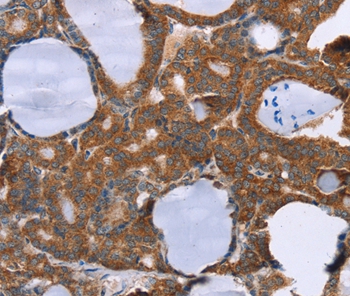

Anti-PITPNM1 Antibody
A45141
ApplicationsImmunoHistoChemistry
Product group Antibodies
Overview
- SupplierAntibodies.com
- Product NameAnti-PITPNM1 Antibody
- Delivery Days Customer7
- ApplicationsImmunoHistoChemistry
- CertificationResearch Use Only
- ClonalityPolyclonal
- Concentration2 mg/ml
- ConjugateUnconjugated
- HostRabbit
- Scientific DescriptionRabbit polyclonal antibody to PITPNM1
- UNSPSC12352203
Related products
Product group Antibodies
Anti-PITPNM1 Antibody144-60167
ApplicationsWestern Blot
TargetPITPNM1
- SizePrice
Product group Antibodies
PITPNM1 AntibodyCSB-PA01995A0RB
ApplicationsELISA
ReactivityHuman
TargetPITPNM1
- SizePrice
Product group Antibodies

PITPNM AntibodyABX430654
ApplicationsFlow Cytometry, ImmunoFluorescence, ELISA, ImmunoCytoChemistry
- SizePrice

Product group Antibodies
Anti-PITPNM1 AntibodyHPA060227
ApplicationsWestern Blot, ImmunoCytoChemistry, ImmunoHistoChemistry
ReactivityHuman
TargetPITPNM1
- SizePrice

Product group Antibodies
Anti-Nir2/PITPNM1 Antibody Picoband(r)A08128-2-CARRIER-FREE
ApplicationsFlow Cytometry, Western Blot, ELISA
TargetPITPNM1
- SizePrice
Product group Antibodies

Goat anti-PITPNM / PITPNM1EB06327
ApplicationsFlow Cytometry, ImmunoFluorescence, ELISA
TargetPITPNM1
- SizePrice
Product group Antibodies
Anti-PITPNM1Y058934
ApplicationsWestern Blot, ELISA, ImmunoHistoChemistry
- SizePrice
Product group Antibodies
Anti-NIR2 AntibodyA83281
ApplicationsFlow Cytometry, ImmunoFluorescence, ELISA
- SizePrice