

Anti-Prion protein [mab 132]
Ab03222-23.0
ApplicationsFlow Cytometry, ImmunoFluorescence, Western Blot, ELISA, Other Application
Product group Antibodies
ReactivityMouse
TargetPRNP
Overview
- SupplierAbsolute Antibody
- Product NameAnti-Prion protein [mab 132]
- Delivery Days Customer7
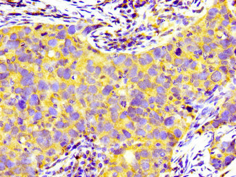
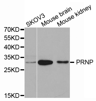
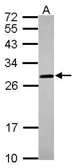
- Application Supplier NoteThe antibody was employed for detection of PrP in N2a-3 cells, ScN2a-3-22L cells, N2a-3 cells infected with the Chandler strain (ScN2a-3-Ch) and in GT1-7 cells persistently infected with the 22L strain (ScGT1-7-22L) in immunofluorescence analysis. Fluorescent signals in uninfected cells remained at a background level, while granular signal were detected in infected cells (Yamasaki et al., 2012; PMID: 22090211). The specificity of the original format of the antibody was confirmed by ELISA analysis (Kim et al., 2004; PMID: 15003861). The antibody could distinguish between prion-infected cells (ScN2a-3-22L) from uninfected cells (N2a-3) in a cell-based ELISA. The cell-based ELISA was successful also with ScN2a-3-Ch and ScGT1-7-22L. (Shan et al., 2016; PMID: 27565564). The antibody reacted with denatured PrPC and PrPSc in western blot analysis. The antibody reacted with mouse ovine and bovine PrP Sc in western blot analysis (Kim et al., 2004; PMID: 15003861). The antibody genetically conjugated with enhanced green fluorescent protein (EGFP) at the C-terminus of the heavy chain was used for the direct immunostaining of PrPSc in ScN2a-3-22L by IFA (Yamasaki et al., 2014; PMID: 25181483). The antibody was used to detect PrPSc in neurons and glial cells from the brains of prion-infected mice by flow cytometry (Yamasaki et al., 2017; PMID: 29046463). Surface plasmon resonance and ELISA analysis revealed that the binding of monovalent recombinant Fab antibody was significantly weaker than bivalent recombinant IgG antibody, indicating that the bivalent binding is required for the efficient binding to the epitope. The binding kinetics of mono and bivalent antibody to MoPrP was measured using SPR, with KD of 5.5 nM for the Fab fragment and 57 pM for IgG1 (Suzuki et al., 2019; PMID: 31170247).
- ApplicationsFlow Cytometry, ImmunoFluorescence, Western Blot, ELISA, Other Application
- Applications SupplierELISA; WB; IF; FC; SPR
- CertificationResearch Use Only
- ClonalityMonoclonal
- Clone IDmab 132
- Gene ID5621
- Target namePRNP
- Target descriptionprion protein (Kanno blood group)
- Target synonymsASCR, AltPrP, CD230, CJD, GSS, KURU, PRIP, PrP, PrP27-30, PrP33-35C, PrPc, p27-30, major prion protein, alternative prion protein, CD230 antigen, prion-related protein
- HostRabbit
- IsotypeIgG
- Protein IDP04156
- Protein NameMajor prion protein
- Scientific DescriptionThis chimeric rabbit antibody was made using the variable domain sequences of the original Mouse IgG1 format for improved compatibility with existing reagents assays and techniques.
- ReactivityMouse
- Reactivity SupplierMouse
- Reactivity Supplier NoteThe original antibody was generated by immunizing PrP gene-ablated mice with PrP.
- Storage Instruction-20°C,2°C to 8°C
- UNSPSC41116161
Related products
Product group Antibodies
PRNP AntibodyCSB-PA018739LA01HU
ApplicationsImmunoFluorescence, ELISA, ImmunoHistoChemistry
ReactivityHuman
TargetPRNP
- SizePrice
Product group Antibodies

Anti-Prion protein [mab 132]Ab03222-1.1
ApplicationsFlow Cytometry, ImmunoFluorescence, Western Blot, ELISA, Other Application
ReactivityMouse
TargetPRNP
- SizePrice
Product group Antibodies
Anti-PRNP AntibodyA30589
ApplicationsWestern Blot, ImmunoHistoChemistry
ReactivityHuman, Mouse, Rat
- SizePrice
Product group Antibodies

PRNP / PrP / Prion AntibodyLS-C830371
ApplicationsELISA, ImmunoHistoChemistry
ReactivityHuman, Mouse, Rat
TargetPRNP
- SizePrice
Product group Antibodies

ApplicationsWestern Blot, ELISA
ReactivityBovine, Human, Porcine
TargetPRNP
- SizePrice

Product group Antibodies
Anti-PRNP AntibodyHPA042754
ApplicationsImmunoHistoChemistry
ReactivityHuman
TargetPRNP
- SizePrice
Product group Antibodies
ApplicationsImmunoPrecipitation, Western Blot, ImmunoCytoChemistry, ImmunoHistoChemistry
ReactivityMouse, Rat
TargetPRNP
- SizePrice

Product group Antibodies
Anti-Prion protein PrP/PRNP Antibody Picoband(r)PB9783-CARRIER-FREE
ApplicationsWestern Blot, ImmunoHistoChemistry
ReactivityHuman, Mouse, Rat
TargetPRNP
- SizePrice
Product group Antibodies
Prion Protein (PrP) antibodyGTX101063
ApplicationsWestern Blot, ImmunoHistoChemistry, ImmunoHistoChemistry Paraffin
ReactivityHuman, Mouse
TargetPRNP
- SizePrice