

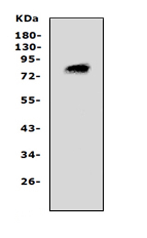
Western blot analysis of PCSK4 using anti-PCSK4 antibody (PA2086). Electrophoresis was performed on a 5-20% SDS-PAGE gel at 70V (Stacking gel) / 90V (Resolving gel) for 2-3 hours. The sample well of each lane was loaded with 50ug of sample under reducing conditions. Lane 1: rat brain lysates. After Electrophoresis, proteins were transferred to a Nitrocellulose membrane at 150mA for 50-90 minutes. Blocked the membrane with 5% Non-fat Milk/ TBS for 1.5 hour at RT. The membrane was incubated with rabbit anti-PCSK4 antigen affinity purified polyclonal antibody (Catalog # PA2086) at 0.5 μg/mL overnight at 4°C, then washed with TBS-0.1%Tween 3 times with 5 minutes each and probed with a goat anti-rabbit IgG-HRP secondary antibody at a dilution of 1:10000 for 1.5 hour at RT. The signal is developed using an Enhanced Chemiluminescent detection (ECL) kit (Catalog # EK1002) with Tanon 5200 system. A specific band was detected for PCSK4 at approximately 83KD. The expected band size for PCSK4 is at 83KD.
Western blot analysis of PCSK4 using anti-PCSK4 antibody (PA2086). Electrophoresis was performed on a 5-20% SDS-PAGE gel at 70V (Stacking gel) / 90V (Resolving gel) for 2-3 hours. The sample well of each lane was loaded with 50ug of sample under reducing conditions. Lane 1: rat brain lysates. After Electrophoresis, proteins were transferred to a Nitrocellulose membrane at 150mA for 50-90 minutes. Blocked the membrane with 5% Non-fat Milk/ TBS for 1.5 hour at RT. The membrane was incubated with rabbit anti-PCSK4 antigen affinity purified polyclonal antibody (Catalog # PA2086) at 0.5 μg/mL overnight at 4°C, then washed with TBS-0.1%Tween 3 times with 5 minutes each and probed with a goat anti-rabbit IgG-HRP secondary antibody at a dilution of 1:10000 for 1.5 hour at RT. The signal is developed using an Enhanced Chemiluminescent detection (ECL) kit (Catalog # EK1002) with Tanon 5200 system. A specific band was detected for PCSK4 at approximately 83KD. The expected band size for PCSK4 is at 83KD.
Anti-ProProtein Convertase PC4 Antibody

PA2086
Overview
- SupplierBoster Bio
- Product NameAnti-ProProtein Convertase PC4 Antibody
- Delivery Days Customer9
- Applications SupplierWB
- CertificationResearch Use Only
- ConcentrationAdd 0.2ml of distilled water will yield a concentration of 500ug/ml.
- Estimated PurityImmunogen affinity purified.
- Protein IDP29121
- Protein NameProprotein convertase subtilisin/kexin type 4
- UNSPSC12352202